
Postpartum hemorrhage: Solutions to 2 intractable cases


A stepwise approach to bleeding caused by persistent uterine atony and placental abnormalities
IN THIS ARTICLE
In a case series, Nishijima and colleagues10 described successful removal of the adherent placenta in 2 patients under direct visualization by inverting the uterus through a large midline uterine incision.
Kayem and colleagues11 described ligation of the umbilical cord close to the placental insertion, with the placenta left in the uterus. They reviewed the records of all patients with the diagnosis of placenta accreta during 2 time frames:
- when management involved immediate hysterectomy
- when management was conservative with the placenta left in utero.
During conservative management, 3 of 20 patients underwent hysterectomy—1 at the patient’s request, 1 at the time of delivery due to hemorrhage, and 1 for bleeding on postoperative day 26 due to endometritis. The rates of disseminated intravascular coagulation and transfusion were lower during conservative management. Two women who underwent conservative management had subsequent successful pregnancies. One of these patients had 2 subsequent pregnancies, both complicated by placenta accreta that was again managed conservatively.
Long-term morbidity of conservative management is unclear
Because of the small number of cases reported, long-term morbidity and mortality rates due to hemorrhage or infection are unknown. Therefore, conservative management should be undertaken with extreme caution! Patients who have completed childbearing should be managed by hysterectomy.
In some cases, conservative management of placenta accreta may serve as a temporizing measure to allow for transfer to a higher level of care when immediate postpartum hysterectomy is not safe or feasible.
When placenta percreta involves the urinary bladder, conservative management may be the only safe option because of the irreparable harm that could occur to the lower urinary tract during hysterectomy.12 (Imagine the urinary tract injuries that could occur during hysterectomy for the patients in FIGURE 3 A and B!)
In these cases, conservative management involves delivering the fetus through a classical uterine incision, ligating the umbilical cord close to the placental insertion, and closing the uterine incision. Avoid attempts to remove the placenta. Give the patient a broad-spectrum prophylactic antibiotic such as amoxicillin/clavulanic acid for 10 days.11 Assess the patient weekly for 6 weeks with ultrasonography, clinical examination, and a white blood cell count with differential (to assess for signs of infection). It may take 6 to 10 months for the placenta to be entirely reabsorbed.
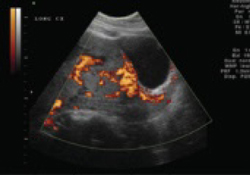
FIGURE 3A Bladder wall involvement
This ultrasonographic image shows placenta percreta involving the posterior bladder wall.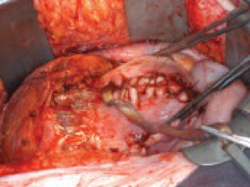
FIGURE 3B Bladder wall involvement
Placenta percreta that has eroded through the entire lower uterine segment and into the bladder. Photo from Stoehr E, Stitely M. Placenta percreta diagnosed antenatally with magnetic resonance imaging. The Female Patient. 2005;30:37–39. Used with permission.
Not all hospitals have the facilities or blood-bank capacity to manage hemorrhagic catastrophes, so temporizing measures to stabilize the patient may be necessary, followed by transfer to a tertiary center.
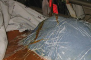
Temporary abdominal closure is a useful strategy for uncontrollable intra-abdominal hemorrhage or coagulopathy. It involves packing the bleeding site with sterile laparotomy pads and sealing it with an occlusive dressing.
To begin, place a sterile x-ray cassette cover into the peritoneal cavity to cover any exposed bowel. Then place moist sterile towels over the cassette cover and any exposed subcutaneous tissue. Place 2 suction drains on top of the towels. Cover the towels and drains with an occlusive adhesive dressing such as an Ioban (3M Healthcare, St Paul, Minn). Attach the drains to wall suction to achieve temporary abdominal closure (see photo).17
Transfer the patient to an intensive care unit for warming, fluid and blood replacement, and correction of acidosis and coagulopathy. Once the acidosis and coagulopathy are reversed, take the patient back to the operating room for removal of the packing and abdominal closure.
If intensive care facilities are unavailable, the patient can be transferred to a tertiary care center following temporary closure.
Manage blood replacement
Few community hospitals keep a large reserve of blood products in stock. A massive obstetric hemorrhage can rapidly deplete blood-bank stores and necessitate transferring the patient or obtaining products from other hospitals or facilities. For this reason, the blood bank should be notified of postpartum hemorrhage as soon as possible, and the possibility of using emergency-release, type-specific blood should be discussed with blood-bank medical personnel.
When to transfuse red blood cells
Administer packed red blood cells (RBCs) if the hemorrhage is profuse and ongoing or if the patient is hemodynamically unstable. Each unit of packed RBCs increases the hematocrit by approximately 3% and raises the hemoglobin level by approximately 1 g.
If coagulopathy is suspected, or the patient has received more than 6 to 8 U of packed RBCs, consider transfusing fresh frozen plasma.


