Bowel Resection With Invasive Endometriosis


Standby status also allows the gynecologist to be more aggressive because rectal wall injury can be corrected at the time of surgery. Flexible sigmoidoscopy always should be performed at the end of any endometriosis operation in which bowel proximity is encountered. By submerging the bowel under water and inflating air via the sigmoidoscope, the presence of air bubbles often will identify a missed bowel injury.
Segmental resection usually involves no more than 5-6 cm of the rectum. We do, however, extend the resection a bit proximally if the patient has a history of chronic constipation. Resecting more of the rectum and sigmoid colon to straighten out the left side of the large bowel and shorten the overall length will better alleviate the patient’s constipation symptoms. Combined with improvement in the patient’s defecation-related symptoms associated with the endometriosis, patient satisfaction regarding the elimination of constipation symptoms is often quite high.
Basic Surgical Technique
Rectal resection for deep endometriosis is comparable to resection of a T4 rectal cancer (one that has invaded outside the rectal wall), except that in the case of endometriosis, we typically are treating young, otherwise healthy patients. In these patients, the risk of complications – mainly, the risk of a permanent colostomy – is all the more concerning. It is important that patients understand the risk and benefits of the surgery and that the colorectal surgeon has the proper expertise for such a technically demanding, risky operation.
The operation is performed in a modified lithotomy position. A laparoscopic or hand-assisted laparoscopic approach can be used. We have performed both techniques, but find a hand-assisted laparoscopic approach faster. A robotic-assisted approach also is being developed.
Depending upon the type of camera used, a 5- or 10-mm port is placed in the umbilicus. The only other ancillary ports needed are 5-mm dissecting ports placed in the right and left lower quadrants. A mini-Pfannenstiel incision is needed to remove the rectal specimen. By extending this incision 3 centimeters, a hand port for hand-assisted laparoscopic surgery can be placed.
Surgery is initiated by the gynecologic surgeon, who resects endometriosis off all nongastrointestinal organs. Endometriosis involving the colon and rectum is left intact. If indicated, hysterectomy and salpingo-oophorectomy is performed at this time.
Next is the laparoscopic colorectal portion of the surgery. First, the inferior mesenteric artery is ligated at the root of the aorta so that various collateral vessels within the marginal branches and Riolan’s arch are not sacrificed. Usually, this ligation alone will adequately free up the sigmoid colon enough for a tension-free anastomosis. If the sigmoid colon still cannot be lowered into the rectum without undue tension, we also will ligate the rectal tributary of the inferior mesenteric vein, one of the two main tributaries of the mesenteric vein.
The remainder of the mobilization involves dissecting along the White line of Toldt until the colon falls freely into the rectum. Rarely will we need to mobilize the splenic flexure of the colon to achieve adequate length.
With the left side of the colon freely mobilized, we turn our attention to the pelvis and subsequent rectal dissection. We do not remove the lesion from the rectum, since we have already confirmed that the lesion is firmly attached to the rectal wall. The endometriosis is removed en bloc with the rectum, similar to what is done for rectal cancer.
While the lateral and posterior dissection of the mesorectum can be easily done laparoscopically or robotically, we believe the anterior dissection of the rectum – removal of the endometriosis off the posterior aspect of the vagina while leaving it attached to the rectum – is more easily performed using a hand-assisted laparoscopic approach or even a hybrid open approach through the mini-Pfannenstiel incision.
Dissection is carried out distally until a soft, normal section of rectum is identified. At least 2 cm of normal rectum is needed for a safe anastomosis.
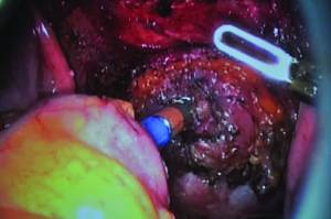
Endometriosis involving the bowel usually appears as a white fibrotic, submucosal mass and feels similar to invasive rectal cancer. The difference, of course, is that rectal cancer is mostly intraluminal, whereas endometriosis usually originates outside the bowel wall and invades inward. Occasionally, one will find "chocolate"-filled cysts within the endometriotic mass, but this is rare.
Endometriosis with bowel involvement is typically anterior to the rectum and posterior to the vagina, but lesions posterior to the rectum have been found, which would denote a nonanatomical spreading distribution.