Failure to thrive


A 13-month-old girl is anemic and not gaining enough weight. How would you proceed with her care?
FIGURE 3
Rachitic rosary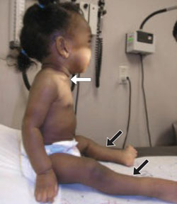
The patient had a developing rosary, most pronounced at the sternoclavicular joint (white arrow) and early saber deformity of the shin (black arrows). Frontal bossing and swollen wrists and ankles may also be seen in patients with rickets.
Not enough vitamin D
The most common cause of rickets is a deficiency of vitamin D, a substance physiologically necessary to produce concentrations of calcium and phosphate adequate for proper bone mineralization. Vitamin D is produced in the skin in the presence of sunlight and can also be ingested in supplements and certain foods.
Vitamin D deficiency may result from reduced sunlight exposure, inadequate dietary intake, malabsorption, or a combination of these factors.1 Rickets may also be caused by medications that alter absorption or secretion of phosphate and calcium, including antacids, anticonvulsants, corticosteroids, and loop diuretics. Various disease states, such as Crohn’s disease, pancreatic disease, biliary disease, gastrointestinal loops and fistulae, cirrhosis, chronic renal disease, and mesenchymal tumors, may also alter absorption and metabolism of these ions.
How much sunshine does a baby need?
It doesn’t take a great deal of sunlight exposure to provide adequate supplies of vitamin D. An infant wearing only a diaper will get enough vitamin D from half an hour per week of sun exposure. A fully clothed infant needs 3 hours. But children with dark skin, like this African baby, need more time in the sun. And if parents follow current anticipatory guidance about protecting children from overexposure to the sun and slather on the sunscreen, vitamin D synthesis decreases by more than 95%.2
Vitamin D in the diet
Consuming adequate quantities of vitamin D is difficult, unless the diet includes fortified foods or vitamin supplements. Current recommendations for daily intake are 400 IU per day for all infants, children, and adolescents.3 But the average daily intake by adults in North America from sources such as fish, eggs, and butter or margarine is only 50 to 100 IU.3
Infants born to vitamin-D replete women have an 8- to 12-week store of vitamin D at birth, but breastfeeding does not ensure the baby is getting adequate amounts of vitamin D, even if the mother’s vitamin D status is adequate. Human milk from vitamin D-replete women has a vitamin D concentration of only 25 IU per liter, far below the recommended daily intake of 400 IU.3
How deficient is this baby?
Vitamin D deficiency may be categorized as mild, moderate, or severe. Calcidiol is the next-to-last step in the metabolism of vitamin D and is used as a marker because it is easier to measure than the concentration of calcitriol, the final step. Mild vitamin D deficiency is defined as serum calcidiol concentration of 25 to 50 nmol/L. A serum calcidiol concentration of 12.5 to 25 nmol/L indicates a moderate vitamin D deficiency, and at those levels the incidence of hypocalcemia and rickets increases. Serum calcidiol concentration of less than 12.5 nmol/L, as in the case of the patient presented here, indicates a severe deficiency.4
Not enough calcium
In addition to calcium malabsorption due to inadequate vitamin D levels, hypocalcemia may result from inadequate intake of calcium or from vitamin D-dependent metabolic disorders, of which there are 2 types.
Type I, sometimes known as pseudo-vitamin D-deficiency rickets, is due to defective production of 25(OH)D3-1-α-hydroxylase, an enzyme necessary for the conversion of calcidiol to calcitriol in the kidneys.
Type II, also called hereditary rickets, is rare. It is caused by mutations in vitamin D receptors and the inability of the ligand to bind or stimulate the proper physiologic response. In this condition, laboratory tests may show high levels of calcitriol.
Not enough phosphorus
Vitamin D-resistant rickets, or familial hypophosphatemic rickets, refers to a clinical presentation of rickets that is caused by a hereditary renal wasting of phosphorus at the proximal tubule level. Laboratory evaluation of a child with this condition will show low phosphate levels, normal calcitriol levels, and hypercalciuria. The specific defect that causes this condition is not known. A family history of short stature, orthopedic abnormalities, poor dentition, alopecia, or parental consanguinity may be suggestive of vitamin D-dependent or vitamin D-resistant forms of inherited rickets.
Other causes of hypophosphatemia include inadequate nutritional intake, X-linked hypophosphatemia, generalized tubular disorders such as renal tubular acidosis, Fanconi syndrome, and Dent disease. These disorders may also lead to rickets.
Q: What are the available treatments for rickets? What about prevention?
Replenishing vitamin D
Vitamin D deficiency may be remedied by supplementing anywhere along the metabolic pathway. Dihydrotachysterol (DHT, or D1), the substance in the skin that responds to sunlight, is given as 60,000 IU once, then 6000 IU daily until the rickets are clinically and radiologically resolved. Further downstream, ergocalciferol (D2) may be given as 1000 to 5000 IU daily for 6 to 12 weeks. The dosage is calibrated by age: 1000 IU/d if age <1 month, 3000 IU/d for ages 1 to 12 months, and 5000 IU/d for children older than 12 months. The final form of vitamin D is cholecalciferol (D3). This is typically administered as either 5000 to 10,000 IU daily for 2 to 3 months, or as 600,000 IU in 1 day, divided into 4 to 6 doses.5,6 All of these supplements taste bad and children are often resistant to swallowing them.