Failure to thrive


A 13-month-old girl is anemic and not gaining enough weight. How would you proceed with her care?
A 13-month-old girl arrives at your clinic, referred by the staff at the Women, Infants, and Children (WIC) nutritional center where her parents—recent immigrants from Africa—go for food supplements. The baby is bundled up in layers of clothing, even though it’s a relatively mild winter day. The father carries her into the examining room and undresses her. The child is tiny and dark-skinned, with curly hair painstakingly divided into little bunches. The parents seem caring, loving, and not particularly worried. They tell you the nurse at the WIC center thought their baby was not gaining enough weight and advised them to bring the baby to you. The referral note from WIC says hemoglobin levels found on routine blood test were low. You list the presenting complaint as anemia.
Q: What are some of the etiologies for anemia in a child this age? What strategies would you use to narrow down the cause?
Additional medical history
- The birth history is unremarkable, with neither antepartum nor postpartum complications.
- At her 6-month well-child checkup, neither the child’s physician nor her parents expressed any concerns about her development. Her parents received the routine anticipatory guidance at that visit, including advice on breastfeeding, vitamin supplementation, vaccination, and care of minor illnesses.
- She hasn’t been in for a well-child visit since then, but she has been seen for an upper respiratory infection and a bout of gastroenteritis. Her parents have not been worried about her health.
- The parents tell you the baby doesn’t sleep soundly, scratches her skin in her sleep, and cries a lot.
Family and social history
- The parents speak very little English.
- The patient is an only child, and no extended family live in the area.
- Her mother works nights and her father works days, with the parent who is not working caring for her at home.
- Her parents tell you she takes small sips of juice or water, and an occasional bite of noodles. She won’t drink milk at all and refuses any other foods they offer.
Physical examination
- The child is in no acute distress. She is afebrile, and her vital signs are appropriate for her age.
- Height is 27½ inches, weight 15 lb, 15 oz, placing her at less than the 5th percentile for height and weight for her age—a regression from the 50th percentile she showed at earlier visits.
- Head and neck exam reveals mild frontal bossing and prominent sternoclavicular joints. There is no adenopathy or thyromegaly.
- Heart and lung exam are normal.
- Abdomen is soft, nontender, nondistended, with bowel sounds present.
- There is slight bowing of the lower extremities and puffiness around the wrists and ankles. When you ask her father to have her stand on the examining table, you see that she needs support to do so (FIGURE 1).
Q: What is your clinical diagnosis, and what tests will you order?
FIGURE 1
The patient, not weight-bearing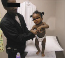
The 13-month-old patient has slight bowing of the lower extremities and puffiness around the wrists and ankles. She needs support to stand.
Laboratory results
- Hemoglobin, 9.9 mg/dL
(normal: 10.4-12.4 mg/dL) - Mean corpuscular volume (MCV), 74 fL
(normal: 70-86 fL) - Alkaline phosphatase, 3417 U/L
(normal: 115-460 U/L) - Vitamin D (calcidiol), <7 nmol/L
(normal: 60-108 nmol/L) - Calcium, 9.1 mg/dL
(normal: 8.8-10.8 mg/dL) - Comprehensive metabolic panel, liver transaminases, and thyroid-stimulating hormone levels are all normal
- Parathyroid hormone level, 101 pg/mL
(normal: 10-55 pg/mL).
Radiologic findings
- X-ray shows slight saber deformity of the femurs and broadening of the epiphyses of the forearm (FIGURE 2).
FIGURE 2
Forearm x-ray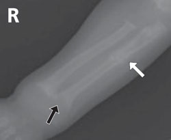
The patient’s x-ray shows a widening of the epiphyses (black arrow) and pseudofracture (white arrow).
Can this be rickets?
Here is a child with a history of poor growth and peculiar eating habits. Her legs are bowed and her wrists seem swollen. She does not stand or walk, and refuses to bear weight on her legs. She is anemic, and the lab tests you’ve ordered show abnormal vitamin D, alkaline phosphatase, and parathyroid hormone levels. All of this suggests a diagnosis of rickets.
Causes of rickets
Rickets is the result of abnormal mineralization of bone and cartilage in growing children. The analogous condition in adults whose epiphyseal plates have closed is osteomalacia. Clinical rickets typically presents with the constellation of signs and symptoms listed in the box and depicted in FIGURES 1, 2, and 3.
Clinical findings
- Apathy, listlessness, and poor growth
- Muscle cramps, weakness, hypotonia, numbness, paresthesias, tetany, and seizures
- Pot belly and waddling gait
- Soft, misshapen head with widened sutures and frontal bossing
- Delayed eruption of teeth
- Rachitic rosary (see below)
- Harrison’s groove (indentation at point of insertion of diaphragm, due to the pull of the diaphragm against the softened lower ribs)
- Bowed limbs and swollen joints
Radiologic findings
- Epiphyses widened and flared with irregular, “cupped” epiphyseal-metaphyseal junctions; involvement of the costrochondral junctions produces a row of beadlike prominences often called the “rachitic rosary”
- Long bones bowed (“saber shaped”), with indistinct cortices
- Pseudofractures (also called Looser’s zone or Milkman’s fractures), often found on the concave side of femoral neck, pubic rami, ribs, clavicles, and lateral aspect of scapulae
- Pathologic fractures
Laboratory findings
- Creatinine: Normal value excludes renal insufficiency as etiology
- Liver enzymes: Normal value excludes liver disease as etiology
- Phosphorus: Normal phosphorus and parathyroid hormone make a diagnosis of rickets unlikely
- Parathyroid hormone: Elevated in hypocalcemic rickets (>55 pg/mL)
- Calcium, total and ionized with albumin: May be normal in rickets
- Alkaline phosphatase: Usually markedly increased over the age-specific reference range in rickets
- Urinary calcium level: Usually decreased in rickets (<50 mg/d, depending on calcium content of diet)
- Calcidiol level: May be low in rickets (<50 nmol/L [20 ng/mL])
- Calcitriol level: Usually normal in rickets, because of its short-half life (60-108 nmol/L [25-45 pg/mL])