Noninvasive vulvar lesions: An illustrated guide to diagnosis and treatment


Dystrophies, vulvodynia, and other noncancerous lesions
IN THIS ARTICLE
Clinical appearance
Macroscopically, VIN lesions are often multiple and appear slightly raised and papular. Hyper- or pseudopigmented lesions are seen in about 30% of cases.
A confluence of VIN can create an appearance of diffuse plaques. In VIN, as for CIN, carcinoma in situ involves a full-thickness abnormality.
Terminology
If the abnormal parabasal layer extends to one half to two thirds of the epithelium, moderate dysplasia is present. A lower degree of involvement is called mild dysplasia, and full-thickness involvement is severe dysplasia or carcinoma in situ.
High rate of persistence, recurrence
Herod and colleagues9 reported on a 15-year follow-up of VIN and found that disease persisted or recurred in 48% of women managed surgically, and 7% of patients progressed to frankly invasive carcinoma. These investigators used the following classification system:
Joura et al10 strongly suggested that the incidence of this condition is increasing, especially in women prior to the 7th decade of life. Whether this increase is due to better recognition or a true rise in prevalence has been debated.
Diagnostic strategies
Application of 5% acetic acid to the vulvar skin will, after 3 to 5 minutes, allow areas of involvement to be readily seen with a handheld magnifying glass. Colposcopy can be used, but is slower and not as efficient as a simple magnifying lens. Most clinicians have abandoned the use of toluidine blue to identify multicentric lesions, since acetic acid appears to be less cumbersome and just as efficient.
White or pseudopigmented lesions (FIGURE 5) can be seen anywhere on the vulva and represent 2 of the 3 presentations of VIN. Pink lesions (FIGURE 5) are usually seen on moist surfaces near mucous membranes, whereas the white or pseudopigmented lesions are usually seen on the drier, hair-bearing areas of the vulva. Biopsy reveals similar histology.
The cause of the pseudopigmented lesions is unclear, but is unrelated to a disturbance of the melanin cells. As with other vulvar lesions, biopsy is essential for a correct diagnosis.
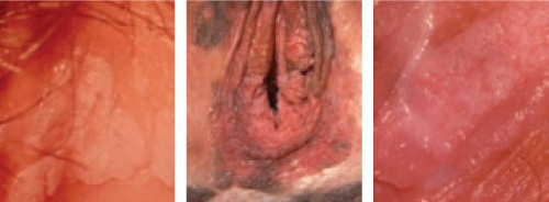
FIGURE 5
VIN III lesions: A trio of presentations
White lesion.
Pseudopigmented lesions.
Pink lesion.
Recommended therapies
VIN is treated by destroying or excising the epithelium involved. When the area is limited in size, simple excision or laser vaporization is preferred. A 2- to 3-mm margin is adequate, because these lesions have sharp borders.
Laser therapy warrants extra care because the epithelium is rarely thicker than 0.5 mm. For this reason, vaporization of the vulva should be limited to the following depths:
These limits will speed healing and prevent the unnecessary destruction of dermis.
Also be aware that the depth of hair-follicle involvement rarely exceeds 1 mm. Vaporization of the full thickness of the dermis will lead to alopecia and vulvar dryness.
When disease is multifocal or confluent, treatment is more challenging. Several decades ago, simple vulvectomy was performed, but the patient was left a sexual cripple.
Since then, laser therapy has been attempted in these cases, and has proved to be effective when the area treated does not exceed 25% of the total area of the vulva. Extensive laser therapy leads to considerable postoperative pain and an unhappy result.
Extensive involvement may necessitate laser treatment by quadrant to achieve the best results. Another option is “skinning vulvectomy” using a skin graft. This procedure, first described by Felix Rutledge,11 requires a 7- or 8-day hospitalization, but allows for complete therapy in 1 session.
Paget’s disease of the vulva
This disease is most frequently seen in the breast nipple (FIGURE 6), where it is usually associated with an underlying infiltrating ductal carcinoma. Extramammary sites include the vulvar, perianal, and axillary regions. The disease has also been seen in the ear canal.
Paget’s disease is an intraepithelial adenocarcinoma of eccrine or apocrine origin.

FIGURE 6
Paget disease: Not just a breast complaint
Paget disease of the nipple with underlying ductal adenocarcinoma.
Vulvar Paget disease: lesion (left) and photomicrograph of the lesion.
Clinical appearance
Paget’s disease of the vulva appears as a superficial, red to pink, velvety, eczematoid lesion (FIGURE 6), which is very pruritic and often associated with exfoliation. Margins are difficult to identify grossly.
On rare occasions, a nodular tumor can be found in the middle of the skin involvement, but in most cases no invasive malignancy is found at extramammary sites on the vulva.
Diagnostic strategies
Microscopic appearance of Paget cells (FIGURE 6) are pathognomonic of the disease.
Older textbooks suggest that 10% to 20% of patients have an associated, underlying, invasive carcinoma of a skin appendage, Bartholin’s gland, urinary tract, or bowel or rectal site, but later experience has not confirmed this. Rather, the likelihood of concomitant invasive disease is much lower than 10%.