Rheumatology has a been a tad slower than other specialties to adopt more advanced imaging modalities, preferring to stick with ultrasound and venturing into MRI. Based on this year’s still image winner in the “Image of the Year” contest at this year’s American College of Rheumatology meeting though, the specialty appears to be embracing innovative new ways of imaging rheumatic diseases.
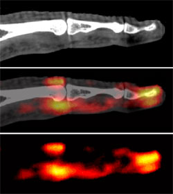
This year’s winner is a combined PET-CT image of the finger joints in patients with psoriatic arthritis. The image was submitted Abhijit Chaudhari, Ph.D. of the UC Davis School of Medicine in Sacramento.
Courtesty ACR and Dr. Chaudhari
This extreme extremity imaging was named ACR's "Image of the Year."
According to Dr. Chaudhari’s poster from the meeting, his group has built an extremity scanner that is capable of sequentially performing 3D positron emission tomography (PET) and fusing the image with a 3D anatomical CT image. In the poster, they reported their initial experience in using this system for assessing metabolic activity in RA, PsA and OA of the hand. Regions of enhancement on PET (F18-FDG) are markers of increased metabolic activity and, in turn, inflammation.
While the technique is still in early trials, the researchers hope that one day they will be able to not only identify the disease but also monitor early response to anti-TNF-alpha therapy in RA and characterize bone remodeling (osteoblastic) activity in early OA.
The best overall submission and category winning submissions from this year’s contest will be published in a future issue of Arthritis & Rheumatism and will be featured in the online Rheumatology Image Bank.
You can read more about this year’s ACR meeting and watch video interviews with key presenters at Rheumatology News.com.