
Two-Toned Toes


Match the diagnosis to the photo by letter
a. Blue toe syndrome
b. Pernio (chilblains)
c. Subungual hematoma
d. Antiphospholipid syndrome
For answers, see next issue; for discussion, go to www.mdedge.com/clinicianreviews/picture
Answers to January “Picture This” (Clinician Reviews. 2018;28[1]:29): 1d, 2c, 3a, 4b
Photos and text for case 1 courtesy of Richard P. Usatine, MD; from Usatine R, Smith M, Mayeaux EJ, et al, eds. Color Atlas of Family Medicine. 2nd ed. New York, NY: McGraw-Hill; 2013:551-556.
To learn more about the Color Atlas of Family Medicine, see: www.amazon.com/Color-Family-Medicine-Richard-Usatine/dp/0071769641/
You can now get the second edition of the Color Atlas of Family Medicine as an app by clicking on this link: usatinemedia.com.
Case presentation and photograph for Cases 2, 3, and 4 courtesy of Cutis. Copyright 2017 Frontline Medical Communications Inc.
2. For the past month, a man in his 50s has had a discolored right foot with increasing tenderness, as well as livedo reticularis on the sole and lateral aspect. He has a history of right-arm arterial thrombosis, multiple deep vein thromboses of the legs, ischemic stroke, atrial fibrillation, peripheral arterial disease, and long-term warfarin treatment. Pulses are palpable on exam.
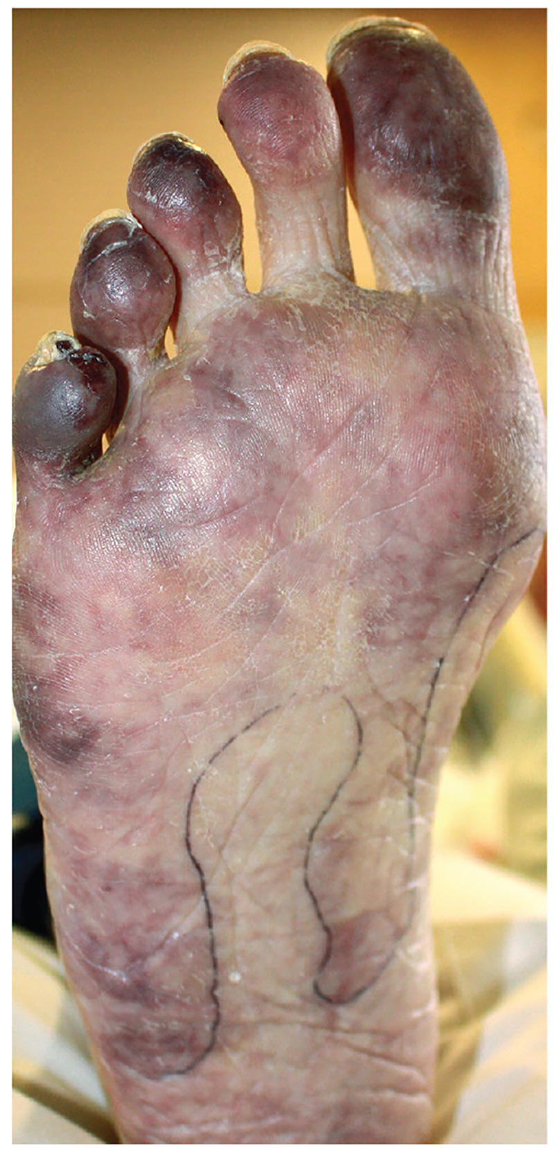
Diagnosis: The patient was diagnosed with antiphospholipid syndrome. He remained on inpatient anticoagulation therapy with fondaparinux and was treated with pulse-dose IV corticosteroids followed by a slow oral taper: daily plasmapheresis for one week, three doses of IV immunoglobulin (0.5 g/kg), and four weekly doses of rituximab (375 mg/m2). His cutaneous findings slowly improved over the next several weeks.
,For more information, see “Cyanosis of the Foot.” Cutis. 2017;100(4):206, 209-210.


