Red eye for the internist: When to treat, when to refer


ABSTRACTWhen a patient presents with redness in the eye, the cause needs to be diagnosed quickly. Some of the diseases that cause redness in the eye can be initially managed by an internist, but others call for quick referral to an ophthalmologist. This article reviews the spectrum of conditions manifesting as a red eye, emphasizing how to differentiate between the benign and the vision-threatening.
KEY POINTS
- Blepharitis, conjunctivitis, corneal abrasion, dry eye, and subconjunctival hemorrhage are benign and can usually be managed initially by an internist, although referral is usually indicated if symptoms persist or progress.
- Patients with corneal bacterial infection, uveitis, scleritis, or acute narrow-angle glaucoma need immediate referral to an ophthalmologist, as do most patients with a red eye who use contact lenses, who have had trauma to the eye, or who have vision changes, severe pain, nausea, vomiting, severe headache, marked purulent discharge, or abnormalities in the cornea or anterior segment.
- Because it is difficult to distinguish between infectious and noninfectious conditions, and because treating infections with corticosteroids alone can have grave consequences, we recommend that internists generally not use topical corticosteroids to treat eye symptoms.
Anterior uveitis
Uveitis is inflammation of the uvea (the pigmented layer between the sclera and retina that includes the iris, ciliary body, and choroid). Anterior uveitis is most commonly idiopathic but can be caused by trauma, secondary to herpes virus infection, or associated with the HLA-B27 antigen.
Acute anterior uveitis presents with pain, photophobia, and blurred vision. Perilimbal (circumcorneal) injection overlies the inflamed ciliary body. The pupil is often constricted and poorly reactive to light. Chronic anterior uveitis, defined as lasting more than 6 weeks, typically presents with gradual vision loss and floaters, rather than with the acute pain or severe redness of acute disease. Anterior uveitis is diagnosed by finding cells and flare in the anterior chamber using a slit lamp.
Refer patients to an ophthalmologist immediately to help avoid visual consequences.22,23 Treatment begins with topical corticosteroid drops and can also include oral corticosteroids or long-term immunosuppresion with corticosteroid-sparing agents.
Nasolacrimal infections
Canaliculitis is an inflammation of the canaliculus, the conduit bringing tears from the eye to the nasolacrimal duct. It presents with mild, unilateral eye redness and a slight discharge that can be expressed from the punctum. It is most commonly caused by Actinomyces israelii infection, but Candida and Aspergillus species can also be involved.
Refer to an ophthalmologist for treatment, which involves mechanically removing the granular material from the canaliculi, combined with probing and irrigating the nasolacrimal system with penicillin G solution.
Dacryocystitis is inflammation of the lacrimal sac (the dilated upper end of the nasolacrimal duct) and is caused by obstruction of the duct. Staphylococcus and Streptococcus species are usually involved. Symptoms include unilateral pain, swelling, and redness over the lacrimal sac at the medial canthus of the eye. Purulent discharge can be expressed from the punctum.
Treatment consists of oral antibiotics with gram-positive coverage followed by surgery to open a passage for drainage from the lacrimal sac into the nasal cavity (dacryocystorhinostomy) once the infection has resolved.24
CONDITIONS NEEDING IMMEDIATE REFERRAL
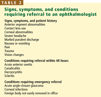
Conditions that require immediate referral to an ophthalmologist can be differentiated from more benign conditions by severe pain or vision loss (Table 2).
Acute angle-closure glaucoma
Patients suffering from an episode of acute angle-closure glaucoma report severe eye pain, seeing halos around lights, headache, nausea, and vomiting. Farsighted people and older people are at greater risk, owing to their eye anatomy. The eyeball is firm to palpation, and the pupil is mid-dilated and poorly reactive to light. The cornea may appear hazy.
Acute angle-closure glaucoma is an emergency and requires immediate lowering of intraocular pressure to avoid permanent vision loss.25
Ocular foreign body
A foreign body lodged in or around the eye causes irritation, redness, and pain. Suspect it in any patient with an appropriate history.
Evert the upper eyelid to search for an occult object and remove any loosely adherent exogenous material on the conjunctiva or sclera. Topical broad-spectrum antibiotic ointments or drops can be started.
Immediately refer any patient with a foreign body that does not dislodge easily for removal and management, or if the patient was working near high-speed objects or with metal on metal (raising the possibility of fragments completely penetrating into the eye).26
Keratitis (corneal inflammation)
Keratitis is inflammation at any level of the cornea.
Herpes keratitis presents with unilateral pain, photophobia, and watering. The most common physical finding is a branching ulcer seen with fluorescein staining under Wood’s lamp illumination. Antiviral treatment with an oral medication (acyclovir [Zovirax] 400 mg five times daily) or topical medication (trifluridine 1% [Viroptic] nine times daily) shortens the course of the disease.27,28 Corticosteroid eye-drops should never be given for epithelial herpetic disease without consulting an ophthalmologist.
Bacterial keratitis threatens sight: infection with a virulent bacterium such as Pseudomonas aeruginosa can cause perforation of the cornea within 48 hours. Patients typically report the rapid onset of pain, photophobia, and decreased vision. Common predisposing risk factors include contact lens use and trauma. Examination reveals infiltration, ulceration, and edema of the cornea, and anterior chamber inflammation. Refer immediately to an ophthalmologist for evaluation and management; delaying treatment can have severe visual consequences.29