Evaluation and management of pituitary incidentalomas


ABSTRACTA surprising number of apparently healthy people harbor unsuspected pituitary tumors, which are being discovered incidentally on computed tomography (CT) or magnetic resonance imaging (MRI) performed for other reasons. The majority can be safely observed; for others, medical therapy or surgical resection is necessary. In this article we outline our approach.
KEY POINTS
- Two key questions that must be answered when a pituitary incidentaloma is discovered are whether it is hormonally active and whether it is causing a mass effect (eg, a visual field defect due to pressure on the optic chiasm).
- Incidentalomas that are not hormonally active and that are not causing a mass effect can generally be managed by watchful waiting.
- Hormonally active prolactin-secreting tumors can be treated with dopamine agonists. Other hormonally active tumors and those that are causing a mass effect should be surgically removed.
- The risks of further tumor growth and of pituitary apoplexy are higher in tumors that are larger when discovered.
IS THERE A MASS EFFECT?
Pituitary macroadenomas can also cause problems via a mass effect. Examples: hypopituitarism, visual field defects (by compressing the optic chiasm), cranial neuropathy (eg, diplopia, eyelid ptosis secondary to lateral extension of the tumor into a cavernous sinus), and headache.
Hypopituitarism
Hypopituitarism can range from deficiency of one pituitary hormone to the loss of all anterior pituitary hormones (panhypopituitarism).
Hypopituitarism from a mass effect is rare in patients with microadenomas, but one or more anterior pituitary hormone deficiencies are found in more than 30% of patients with a pituitary macroadenoma.3,12,14 With some exceptions, including pituitary apoplexy, the loss of pituitary hormone secretion is slowly progressive; symptoms tend to be nonspecific and often are not noticed at first.
Increased intrasellar pressure may play a role in the pathogenesis of hypopituitarism in patients with pituitary masses.15 Blood flow through the portal vessels is decreased, possibly resulting in diminished delivery of hypo-thalamic hormones to pituitary cells or leading to variable ischemia or necrosis of the normal gland, or both.
All patients with a pituitary macroadenoma should undergo a hormonal evaluation to look for pituitary hormone deficiency.
Growth hormone, gonadotropin deficiencies. In general, pituitary hormone deficiencies from an expanding pituitary tumor tend to begin with growth hormone or the gonadotropins (luteinizing hormone and follicle-stimulating hormone), or both.
Low serum testosterone levels in men (estradiol in women) along with normal or low follicle-stimulating hormone and luteinizing hormone levels are consistent with gonadotropin deficiency in men and amenorrheic premenopausal women.
Failure of the follicle-stimulating hormone and luteinizing hormone levels to rise after menopause is also consistent with gonadotropin deficiency. The presence of regular menses almost always indicates a normal gonadotropin axis. In women with irregular menstruation, hormonal evaluation can be challenging for evaluation of the gonadotropin axis and usually is not indicated.
Patients with deficiencies of two or more pituitary axes and low IGF-1 levels can be presumed to have growth hormone deficiency and usually do not need dynamic testing. But when testing is indicated, the growth hormone axis is best evaluated by dynamic testing, using either a growth hormone-releasing hormone/ arginine stimulation test or the insulin tolerance test.
Thyroid deficiencies. As the tumor expands, deficiencies of thyrotropin and adrenocorticotropic hormone (ACTH) secretion may follow those of growth hormone and gonadotropins. In our experience, the thyrotropin axis is usually affected before the corticotropin axis.
To evaluate the thyrotropin axis, the serum thyrotropin level should be measured along with the free thyroxine level or the free thyroxine index. A low free thyroxine level with a low or normal thyrotropin level is consistent with secondary hypothyroidism. It is inappropriate to measure thyrotropin without also measuring thyroxine in a patient with pituitary disorder, since a normal thyrotropin level in a patient with hypopituitarism is not uncommon.
Adrenal insufficiency. The ACTH stimulation test or an early morning (8 am) plasma cortisol level are both reasonable initial tests to evaluate the hypothalamic-pituitary-adrenal axis. An early morning cortisol level lower than 3 μg/dL confirms adrenal insufficiency, while a value higher than 15 μg/dL makes the diagnosis highly unlikely. Cortisol levels in the range of 3 to 15 μg/dL are indeterminate and should be further evaluated by an ACTH stimulation test, which can be performed anytime during the day.
The standard-dose ACTH stimulation test uses an intravenous or intramuscular injection of 250 μg of cosyntropin (Cortrosyn; ACTH 1–24). A normal response is a plasma cortisol concentration higher than 18 μg/dL at 30 minutes.
The sensitivity of the ACTH stimulation test in detecting mild, partial adrenal insufficiency is higher if a lower dose of cosyntropin is used (1 μg intravenously). However, the low-dose test has a higher false-positive rate. In most clinical situations, the 30-minute cortisol value during a standard-dose ACTH stimulation test has a diagnostic accuracy close to that of the low-dose ACTH stimulation test.16 Patients with recent-onset ACTH deficiency (eg, in pituitary apoplexy or within 2 to 4 weeks following pituitary surgery) may have a normal response to the ACTH stimulation test, since their adrenal glands have not undergone sufficient atrophy and still respond to ACTH stimulation.
The insulin tolerance test is considered the gold standard for evaluating the hypothalamic-pituitary-adrenal axis, but it needs to be performed by an experienced clinician and is usually not needed for everyday clinical practice.
Visual field defects
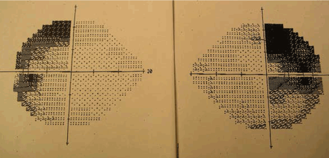
Visual field loss generally begins in the superior temporal fields, which explains why the patient may not notice it at first. Then, with continued growth and compression, vision loss extends into the inferior temporal fields, then into the nasal fields as a late effect.
Because the patient may not notice the visual field defect, formal visual field testing is warranted if the tumor compresses or abuts the optic chiasm. While bitemporal hemianopia is the classic manifestation of chiasmal compression, variable visual field defects may occur depending on which portion of the optic apparatus is involved.