CT imaging for acute aortic syndrome


ABSTRACTAcute aortic syndrome can be due to acute aortic dissection, intramural hematoma, penetrating atherosclerotic ulcer, or unstable thoracic aneurysm. These life-threatening conditions are clinically indistinguishable, often presenting with acute chest pain. Contrast-enhanced, cardiac-gated multidetector computed tomography (CT) is a highly accurate imaging method for determining the cause of acute aortic syndrome.
KEY POINTS
- Acute aortic syndrome typically presents with chest pain in patients with a history of hypertension. In young patients with aortic dissection, one should consider Marfan syndrome and other connective tissue abnormalities.
- Cardiac gating is essential to avoid cardiac motion artifacts when evaluating the aortic root with contrast-enhanced multidetector CT.
- Urgent surgical repair is often necessary, especially for acute aortic dissection and intramural hematoma in the ascending aorta and aortic arch, unstable or ruptured thoracic aneurysm, and symptomatic penetrating atherosclerotic ulcers.
Aortic intramural hematoma
Intramural hematomas are believed to be caused by a spontaneous hemorrhage of the vaso vasorum into the medial layer. They appear as crescent-shaped areas of increased attenuation with eccentric aortic wall-thickening and displacement of intimal calcifications. Hematomas do not enhance after contrast administration, and unlike dissections, they usually do not spiral around the aorta.
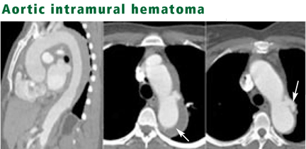
Type B intramural hematomas are typically managed with medical therapy and often regress with time, although they can progress to dissection or aneurysmal formation.
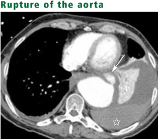
Unstable thoracic aneurysm
Thoracic aneurysms are considered unstable if they are enlarging rapidly, show signs of imminent rupture, or have already ruptured (typically ,the rupture is contained if the patient survives for imaging).
An aortic aneurysm is defined as a permanent dilation at least 150% of normal size, or larger than 5 cm if in the thoracic aorta or larger than 3 cm if in the abdominal aorta. True aneurysms involve all three layers of the aorta and tend to be fusiform; pseudoaneurysms tend to be saccular and often arise after trauma, surgery, or infection. Dilations are more likely to rupture if they grow at least 1 cm per year or measure 6.0 cm or more (if in the ascending aorta) or 7.2 cm (if in the descending thoracic aorta).13
How big the aortic diameter needs to be before invasive treatment—surgery or an endovascular procedure—is indicated depends on the characteristics of the individual patient, and an experienced surgeon should be involved in the decision. Patients are typically treated when a dilation in the ascending aorta reaches 5.5 cm or when one in the descending aorta reaches 6.0 cm; patients with Marfan syndrome should undergo invasive treatment for aneurysms with smaller diameters.14,15
CT signs of imminent rupture include a high-attenuating crescent in the wall of the aorta, discontinuous calcification in a circumferentially calcified aorta, an aorta that conforms to the neighboring vertebral body (“draped” aorta), and an eccentric nipple shape to the aorta.16,17
Penetrating atherosclerotic ulcer
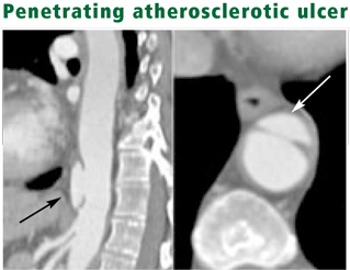
Typical patients are elderly, and many have coexisting atherosclerotic atheromata and aneurysmal disease. Some experts contend that most saccular aneurysms are caused by penetrating atherosclerotic ulcers.19
Surgery to stabilize disease is recommended for a penetrating ulcer that causes acute aortic syndrome, or in patients with hemodynamic instability, aortic rupture, distal embolization, or a rapidly enlarging aorta. For a penetrating ulcer that is found incidentally in a patient without acute aortic syndrome, medical management of risk factors is recommended, with annual follow-up to see if it enlarges.
FUTURE USES OF IMAGING IN PATIENTS WITH ACUTE CHEST PAIN
Multidetector CT systems are undergoing rapid technical advances, including the recently released dual x-ray source multidetector CT scanners and the expected multidetector CTs with 128 to 256 detector rows. These and other developments will improve temporal resolution and decrease radiation exposure. Calcium artifacts—which hinder the evaluation of coronary arteries that contain atherosclerotic calcifications—will be reduced, thereby improving the accuracy of diagnosing acute coronary disease. As clinical knowledge increases based on the experience gained from the new technology, indications for imaging may expand.
CT of the chest performed for aortic disease provides information about other organ systems that may be considered when evaluating the cause of chest pain.20
Evaluating pulmonary artery embolism
Although current contrast-enhanced, cardiac-gated CT of the aorta is not ideal for assessing the pulmonary arteries, it can almost always rule out central pulmonary artery thromboemboli and evaluate the more distal pulmonary arteries in a more limited way.
‘Triple rule-out’ CT
Several institutions now use CT (typically with 64-row scanners) to simultaneously evaluate patients for coronary artery disease, acute aortic syndrome, and pulmonary embolism—or “triple rule-out CT.” The study can be performed with dual intravenous contrast bolus techniques with nongated contrast-enhanced CT of the pulmonary arteries, rapidly followed by cardiac-gated, contrast-enhanced CT of the aorta. The pulmonary arteries can be evaluated on the initial nongated study, and the aorta (including the aortic root) and the coronary arteries are evaluated on the cardiac-gated portion. The timing of the contrast bolus for optimal opacification of the pulmonary arteries and the coronary arteries has yet to be determined.
The usefulness of assessing all three vascular beds with a single study is currently still unclear and the protocol is currently not routinely performed at Cleveland Clinic. Its sensitivity, specificity, and cost-benefit ratio are also unclear and must be determined in prospective clinical trials, which are currently under way.21