ABSTRACTAcute aortic syndrome can be due to acute aortic dissection, intramural hematoma, penetrating atherosclerotic ulcer, or unstable thoracic aneurysm. These life-threatening conditions are clinically indistinguishable, often presenting with acute chest pain. Contrast-enhanced, cardiac-gated multidetector computed tomography (CT) is a highly accurate imaging method for determining the cause of acute aortic syndrome.
KEY POINTS
Acute aortic syndrome typically presents with chest pain in patients with a history of hypertension. In young patients with aortic dissection, one should consider Marfan syndrome and other connective tissue abnormalities.
Cardiac gating is essential to avoid cardiac motion artifacts when evaluating the aortic root with contrast-enhanced multidetector CT.
Urgent surgical repair is often necessary, especially for acute aortic dissection and intramural hematoma in the ascending aorta and aortic arch, unstable or ruptured thoracic aneurysm, and symptomatic penetrating atherosclerotic ulcers.
INTRAVENOUS CONTRAST IMPROVES IMAGING STUDIES
Intravenous contrast is necessary for CT to achieve its high accuracy for diagnosing aortic disease. However, it should be noted that in a CT study without contrast enhancement, an acute intramural hematoma is easily recognized by the higher Hounsfield-unit value of the blood products in the wall in comparison with the flowing blood in the lumen.
Check renal function
Before doing an intravenous contrast study, the serum creatinine level should be checked as a measure of renal function. Generally, if the serum creatinine concentration is less than 2.0 mg/dL and if the patient is hydrated and does not have diabetes, iodinated intravenous contrast can be given safely. If the patient’s serum creatinine level is between 1.5 and 2.0 mg/dL or if he or she is dehydrated or has diabetes, isosmolar iodinated contrast (iodixanol [Visipaque]) may be used.
An alternative that can be used for some patients with contrast allergy is gadolinium chelate, a paramagnetic compound normally used in magnetic resonance imaging. However, it is less radiopaque and much more expensive than iodinated contrast. It should be noted that the US Food and Drug Administration has recently warned that gadolinium contrast is associated with a systemic fibrosing disorder (nephrogenic systemic fibrosis) in patients with poor renal function (usually but not only in patients on dialysis).8 Because of this risk, gadolinium contrast should generally be used only in patients with good renal function who have had a prior serious adverse reaction to iodinated contrast.
Insert a large-gauge intravenous line
Proper intravenous access is needed for contrast injection. To opacify the aorta properly, the contrast must be injected rapidly (3.0 mL/second) using a power injector.
We recommend at least an 18-gauge peripheral intravenous catheter in the forearm or a large-bore central line (an introducer or Hickman catheter). Smaller-gauge intravenous lines (often located in smaller veins such as in the wrist) and most central lines placed without radiographic or surgical assistance (eg, triple-lumen central catheters) cannot safely handle such a rapid rate without infiltration or embolization. Some peripherally inserted central catheters are designed to handle high injection rates and are typically labeled with the injection rate.
USE OF CT IN SPECIFIC ACUTE AORTIC SYNDROMES
Acute aortic dissection
Aortic dissection occurs when a tear in the intimal layer allows blood to enter and accumulate in the medial layer of the aorta, giving rise to a true lumen and a false lumen separated by an intimomedial flap. Dissection is considered to be acute if symptoms have been present for less than 2 weeks.9
Aortic dissections are often complex and can spiral around the aorta. The relationship of the intimomedial flap to the coronary arteries, aortic-arch branch vessels, and visceral branch vessels can be described on contrast enhanced, cardiac-gated CT scans. The true lumen is often smaller and more opacified with contrast than the false lumen; intimal calcification often surrounds the true lumen. Slender areas of low attenuation (“cobwebs”) are occasionally seen in the false lumen. The false lumen also has beaked edges where it meets the true lumen, which usually appears rounder.2,10 The radiologist should state where the dissection begins and ends, determine if target vessel ischemia is evident, and assess for concomitant aneurysmal dilatation of the aorta. Contrast-enhanced, cardiac-gated multidetector CT of the aorta is necessary to properly evaluate the aortic root.
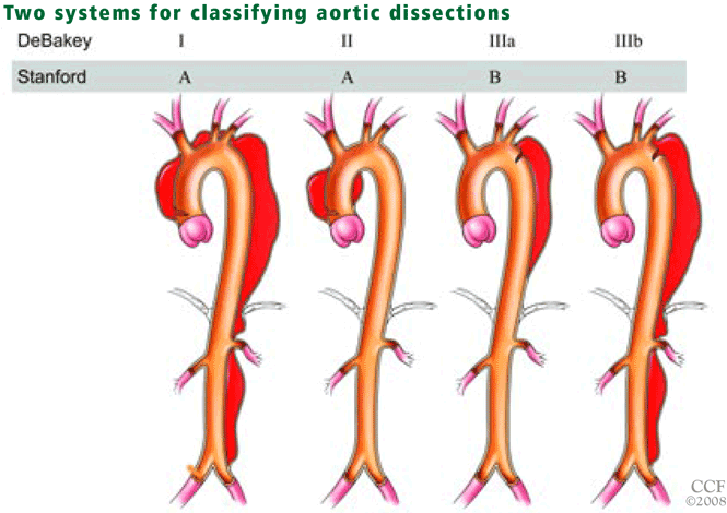
Figure 3. The DeBakey and Stanford systems.
The Stanford system. Two systems exist for classifying the location of aortic dissections: the DeBakey system and the Stanford system (Figure 3). The Stanford system is more clinically useful and uses the following classification:
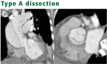
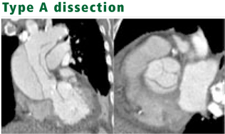
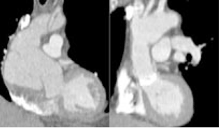
Figure 4. Coronal reformatted image (left) and oblique reformatted image (right) from contrast-enhanced, cardiac-gated computed tomography in a patient with acute aortic syndrome show a type A aortic dissection involving the aortic root, extending around the aortic valve, and aneurysmal dilatation of the aortic root.
Type A dissections involve the ascending aorta and aortic arch, with or without involvement of the descending aorta (Figure 4, Figure 5)
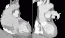
Figure 5. Oblique reformatted images from contrast-enhanced, cardiac-gated CT before (left) and after (right) surgical aortic root repair with aortic valve replacement in a patient who initially presented with acute aortic syndrome and had a type A acute aortic dissection with aneurysmal dilatation of the aortic root.
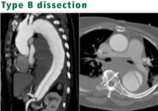
Type B dissections involve the descending aorta beginning distal to the left subclavian artery (Figure 6).11
Figure 6. A coronal reformatted image (left) and an axial image (right) from contrast-enhanced, cardiac-gated CT in a patient who presented with acute aortic syndrome show a type B aortic dissection extending from the aortic arch (distal to the arch vessels) into the abdomen. Hemorrhage from recent rupture is seen in the left and right hemithorax and in the mediastinum (arrow).
Type A acute aortic dissection generally should be surgically repaired immediately to avoid fatal complications such as extension into the pericardium, pleural space, coronary arteries, or aortic valvular ring. It can also cause stroke, visceral ischemia, or circulatory failure.2,11 Without surgery, 20% of patients with type A acute aortic dissection die within 24 hours, 30% within 48 hours, 40% within 1 week, and 50% within 1 month.2 The initial target is the tear in the ascending aorta: typically the aortic root or the ascending aorta or both are replaced and the aortic valve is repaired if indicated (Figure 5). Further aortic repair can often be delayed or may not be needed if the disease does not progress with medical management.
Without surgery, type B acute aortic dissection has a 30-day mortality rate of 10%.2 Patients who develop renal failure, ischemic leg symptoms, or visceral ischemic symptoms with acute aortic syndrome should undergo imaging of the chest, abdomen, and pelvis. Type B acute aortic dissection without end-organ ischemia is typically managed with antihypertensive drugs. Except in patients with Marfan syndrome, only a small minority of type B dissections progress to type A dissections.Urgent aortic repair, often with an endovascular stent graft, is needed if imaging shows visceral vessel occlusion or ischemia, acute vessel thrombosis, or progression of aneurysmal dilatation.