Novel device aims to make cervical cancer screening more accessible


A novel vaginal inserter currently in development at Duke University for use with a miniature colposcope proved feasible for comfortable, speculum-free cervical image capture in a recent study.
Along with allowing for more comfortable – and possibly self-facilitated – cervical cancer screening, the device could have important implications for providing care and improving screening rates in underserved, low-resource communities in the United States and around the world, according to Mercy N. Asiedu, a graduate student research assistant in the Duke University department of biomedical engineering.
Ms. Asiedu is part of a team that has been working to develop effective, low-cost methods for diagnosing cervical and breast cancer, particularly in low-resource regions.
A device such as the integrated inserter and colposcope could potentially be purchased over the counter at a local pharmacy and used for “self checkups,” or used in clinics to facilitate a more comfortable, patient-friendly approach to exams here in the United States, she said in an interview.
“But in other countries, one of the bigger benefits [relates to] cultural factors that deter women from going in for gynecological exams,” Ms. Asiedu said.
Overcoming screening barriers
Invasive cervical cancer ranks as the second most common female cancer in low and middle-income countries and the seventh most common in high-income countries, she and her colleagues wrote in a study published in PLoS ONE (12[5]:e0177782. doi: 10.1371/journal.pone.0177782).
The discrepancies between high- and low-resource areas largely result from the fact that visualization of the cervix via colposcopy using a standard approach requires a highly-trained professional and expensive equipment that is not easily accessible in underserved regions.
But even in the United states, the use of a standard speculum to expand the vaginal canal has been identified as a factor in the resistance of some women to undergo screening. Embarrassment and fear of pain have also been reported as potential barriers to screening.
The ability to self-insert can also help reduce pain associated with the exam, as it allows the user to adjust positioning when there is discomfort, she said.
A speculum alternative
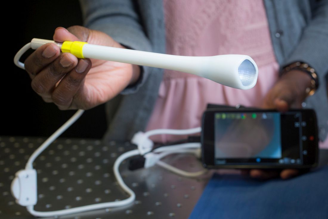
The inserter is a tampon-sized tubular device designed as an alternative to a standard speculum, and is used with the Pocket Colposcope, a miniature pen-sized colposcope also developed by Duke University researchers and validated by physicians worldwide as providing image quality comparable to high-end, state-of-the-art digital colposcopes.
The current rendition of the inserter device has a funnel-like, curved tip measuring approximately 2.6 cm in diameter, a channel for insertion of a 2-megapixel mini-USB camera with LED illumination for the cervical image capture (as opposed to the 5-megapixel camera typically used with the Pocket Colposcope in the setting of standard speculum use), and can connect to a number of devices such as smartphones and laptop computers for image display.
“The curved tip enables easy manipulation of the cervix, especially in cases where the patient has a tilted uterus,” Ms Asiedu and her colleagues wrote in PLoS. “Our design also enables manipulation of the cervix for cervical imaging of women with tilted uteri, a condition that affects about 20% of women and is difficult and painful when using the standard speculum for manipulation.”
Using a custom-made vaginal phantom, the researchers tested various designs and demonstrated that it was able to withstand a range of supine vaginal pressures. In addition, 12 of 15 healthy volunteers achieved adequate cervical image capture after self-insertion of the device, and 14 of the 15 women expressed an overall preference for the inserter over the speculum (based on past experience). All of the volunteers said the inserter was slightly more, or much more, comfortable than a standard speculum, and noted that comfort was a particular benefit.
Prior to self-insertion, the volunteers typically received about 5 minutes of instruction using a pelvic mannequin. Images were captured and displayed on a mobile device once the cervix was in view.
From a patient perspective, the volunteer study “was almost universally positive,” John W. Schmitt, MD, professor of obstetrics and gynecology, as well as global health, at Duke, said in an interview. He conducted the clinical portion of the research.

From the clinician perspective, there is a bit of a learning curve, he added.
“I’ve approached it with what I’d call healthy skepticism,” he said, noting that traditional specula have been around for ages, and are easy to use. “I’ve used them for a long time and I know how, so there’s a learning curve to get it right [with the new device], but as I’ve done more and more, it’s becoming really obvious that this can be used, and ... it’s got real potential to make for a more comfortable internal exam.”
That is particularly true if the size and design can be maintained while also allowing channels to be added to allow solutions, such as acetic acid or Lugol’s solution, to be applied to the cervix to help with visualization – all while keeping the camera in place and allowing the insertion of swabs for obtaining samples from the cervix, he said.