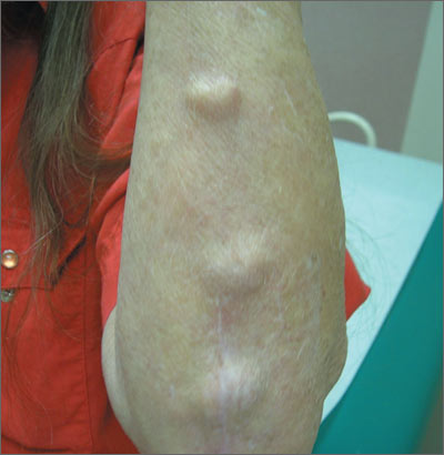
Lumps on forearm


A 48-year-old woman presented to the family physician (FP) with 3 nonpainful lumps on her left forearm that had developed 6 months earlier. The patient was new in town and indicated she’d previously been treated for arthritis. She said she took ibuprofen daily for morning stiffness in her fingers and swelling during the day. On physical exam, the masses were somewhat firm and partially movable.
What's your diagnosis?
The FP recognized that this was a case of rheumatoid arthritis (RA) with rheumatoid nodules. She ordered bilateral hand x-rays and referred the patient to rheumatology. The patient was told that rheumatoid nodules are not harmful and surgery wasn’t recommended. The FP also ordered appropriate blood tests to confirm the impression of RA, which included:
- a rheumatoid factor (RF) test (positive in many connective tissue, neoplastic, and infectious diseases; positive in 70% of RA patients)
- an anticitrullinated protein antibody (ACPA) test (high specificity for RA: Often present before definitive diagnosis can be made; presence predicts arthritis development)
- C-reactive protein (CRP) >0.7 pg/mL, or erythrocyte sedimentation rate (ESR) >30 mm/h
- complete blood count (indicators of RA: normocytic or microcytic anemia, thrombocytosis)
The 2010 American College of Rheumatology/European League Against Rheumatism classification criteria uses a scoring system to identify patients with RA. A score of ≥6/10 meets the criteria.
- joint involvement: 1 large joint (0 points); 2 to 10 large joints (1 point); 1 to 3 small joints with or without large joints (2 points); 4 to 10 small joints with or without large joints (3 points); more than 10 joints with at least 1 small joint (5 points)
- serology: Negative RF and ACPA (0 points); low positive RF or ACPA (2 points); high positive RF or ACPA (3 points)
- acute-phase reactants: Normal CRP and ESR (0 points); abnormal CRP or ESR (1 point)
- duration of symptoms: <6 weeks (0 points); ≥6 weeks (1 point)
Photo and text for Photo Rounds Friday courtesy of Richard P. Usatine, MD. This case was adapted from: Chumley H. Rheumatoid arthritis. In: Usatine R, Smith M, Mayeaux EJ, et al, eds. Color Atlas of Family Medicine. 2nd ed. New York, NY: McGraw-Hill;2013:575-579.
To learn more about the Color Atlas of Family Medicine, see: www.amazon.com/Color-Family-Medicine-Richard-Usatine/dp/0071769641/
You can now get the second edition of the Color Atlas of Family Medicine as an app by clicking on this link: usatinemedia.com