Fleshy tumors


A 44-year-old Hispanic man asked his family physician (FP) if any of his skin tags could be removed. The FP noted many soft fleshy tumors on the patient's skin and a number of light brown patches. The patient said that he started noticing the skin tumors during his teen years.
What's your diagnosis?
| 
|
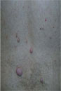
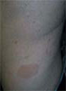
| FIGURE 1 | FIGURE 2 |
This man was given a diagnosis of neurofibromatosis, type 1 (NF-1). He had the typical features of NF-1, including neurofibromas all over his body (FIGURE 1), 8 café au lait spots (FIGURE 2), and axillary freckling.
NF-1 is relatively common: birth incidence is one in 3000 and prevalence in the general population is one in 5000. While it is typically inherited in an autosomal-dominant fashion, up to 50% of cases are sporadic. The diagnosis is typically made during childhood, but it may not be recognized until later in life.
For a diagnosis of NF-1, patients need to have at least 2 of the following:
- >=2 neurofibromas or >=1 plexiform neurofibromas
- >=6 café au lait spots (0.5 cm or larger before puberty, 1.5 cm or larger after puberty)
- axillary or inguinal freckling
- optic glioma
- >=2 Lisch nodules (melanotic iris hamartomas)
- dysplasia of the sphenoid bone or dysplasia/thinning of long bone cortex
- a first-degree relative with NF-1.
This patient’s skin tags were not removed. No intervention was necessary at this time other than recommending yearly visits to the ophthalmologist (to watch for the development of gliomas) and to his family physician to monitor his condition.
Photos and text for Photo Rounds Friday courtesy of Richard P. Usatine, MD. This case was adapted from: Chumley, H. Neurofibromatosis. In: Usatine R, Smith M, Mayeaux EJ, et al, eds. The Color Atlas of Family Medicine. New York, NY: McGraw-Hill; 2009:982-985.
To learn more about The Color Atlas of Family Medicine, see:
• https://www.amazon.com/Color-Atlas-Family-Medicine/dp/0071474641
You can now get The Color Atlas of Family Medicine as an app for mobile devices by clicking this link: