Painful body lesions


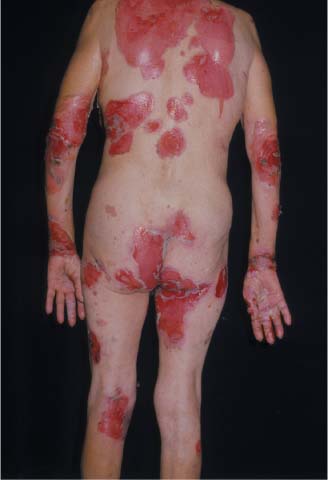
A 62-year-old woman asked her family physician (FP) to take a look at some painful lesions that had developed all over her body 4 weeks earlier. The lesions resembled second-degree burns, but there was no history of exposure to fire or corrosive chemicals. The FP immediately called a dermatology colleague, who suggested that the patient be admitted for IV fluids and intensive treatment. Upon further questioning, the patient admitted to having some skin blisters that peeled off in the previous week. The patient was feeling weak, and had not been eating or drinking much in the previous few days.
What's your diagnosis?
The diagnosis
Two stat shave biopsies—one from the edge of an erosion for hematoxylin and eosion staining, and the other of perilesional skin for direct immunofluorescence—revealed a diagnosis of pemphigus foliaceous.
The large areas of skin erosion impaired the skin barrier function and made this patient similar to a burn patient. The patient was at risk of dehydration and superinfection. As a result, IV fluids were started in the hospital and intake and output were measured. The dermatologist prescribed oral prednisone and topical triamcinolone ointment twice daily for the open erosions.
The patient survived under the care of the dermatologist.
Text for Photo Rounds Friday courtesy of Richard P. Usatine, MD. Photos courtesy of Eric Kraus, MD. This case was adapted from: Mittal S. Pemphigus. In: Usatine R, Smith M, Mayeaux EJ, et al, eds. The Color Atlas of Family Medicine. New York, NY: McGraw-Hill; 2009:794-798.
To learn more about The Color Atlas of Family Medicine, see:
• https://www.amazon.com/Color-Atlas-Family-Medicine/dp/0071474641
The Color Atlas of Family Medicine is also available as an app for mobile devices. See