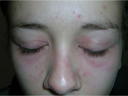
Rash on face


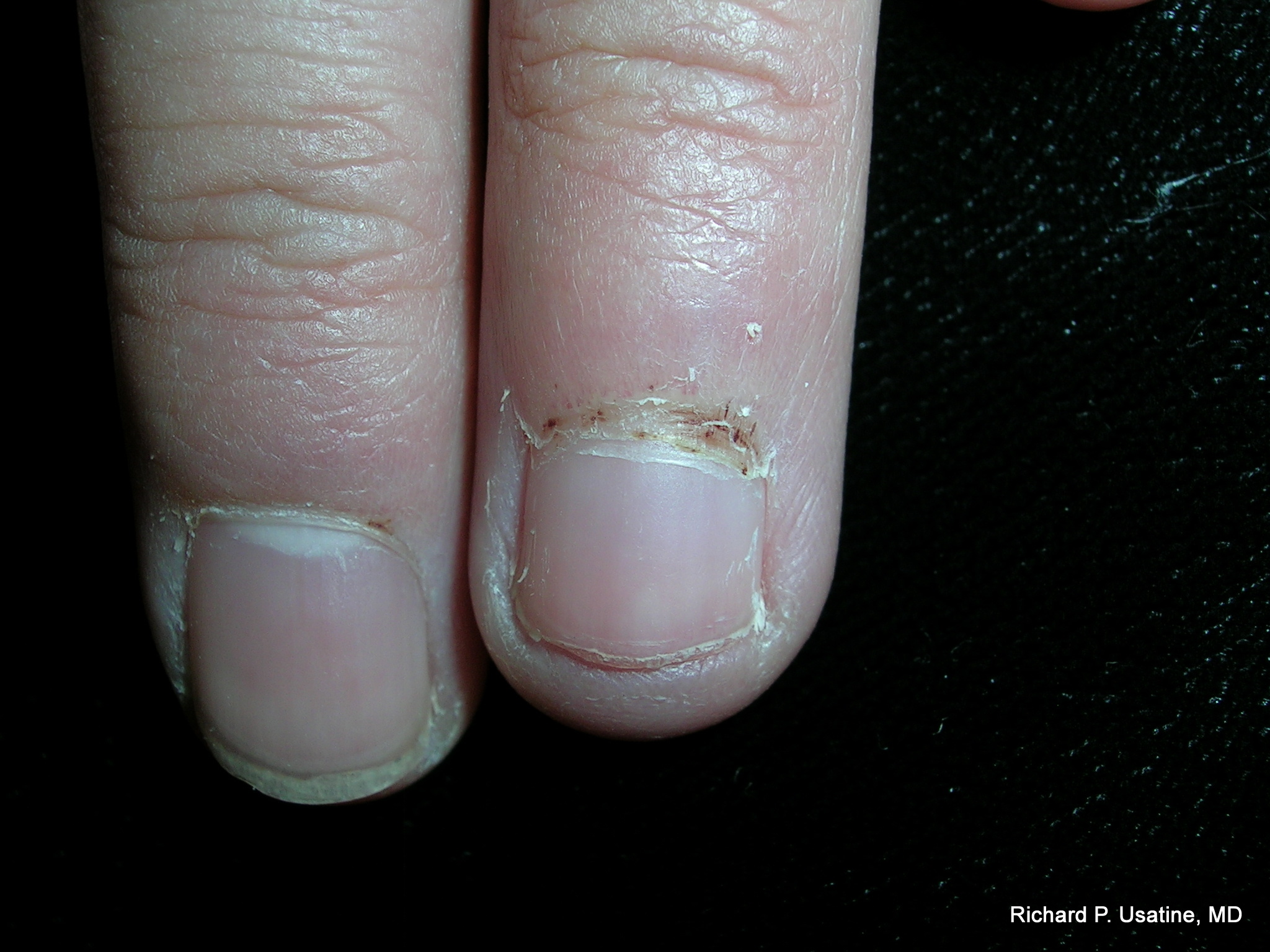
A 19-year-old woman went to see her family physician (FP) about a new rash on her face. She worked as a waitress and commented that the heavy trays she’d been carrying felt a bit heavier lately. She noted that the rash seemed worse on her days off work. The rash was most prominent around her eyes, but seen nowhere else on the body. In looking at her fingers, her cuticles did not appear normal even though her nails were. She had a few papules over the metacarpophalangeal joints of her hand. Examination of her muscle strength was normal, aside from some mild weakness in the deltoid muscles.
What's your diagnosis?
|
|
The patient was given a diagnosis of dermatomyositis, even though muscle weakness was barely present. FIGURE 1 shows a classic heliotrope rash around the patient’s eyes, which was bilaterally symmetrical. The facial rash was getting worse on her days off because she was spending more time in the sun and the rash of dermatomyositis is often worsened by sun exposure. Hand involvement consisted of Gottron’s papules over the finger joints and ragged cuticles (Samitz sign) (FIGURE 2). The patient had normal muscle enzymes. Her diagnosis could almost be called amyopathic dermatomyositis, which is dermatomyositis without muscle involvement.
The physician started the patient on prednisone 60 mg daily and after 2 weeks the rash was almost completely resolved. The patient was also counseled to avoid the sun when possible, and to use sun protection when outside.
The patient was started on 400 mg hydroxychloroquine daily and in 2 months, the prednisone was completely tapered off.
The physician sent the patient to ophthalmology to have a baseline retinal exam at the time the hydroxychloroquine was started. At 6 months, her eye exam remained normal despite the risk of eye problems from the hydroxychloroquine. A year later, the patient had stopped the hydroxychloroquine and was doing well off the medication. Two years later the patient had a relapse and was treated successfully using the same regimen.
Photos and text for Photo Rounds Friday courtesy of Richard P. Usatine, MD. This case was adapted from: Allred A, Usatine R. Dermatomyositis. In: Usatine R, Smith M, Mayeaux EJ, et al, eds. The Color Atlas of Family Medicine. New York, NY: McGraw-Hill; 2009:772-777.
To learn more about The Color Atlas of Family Medicine, see:
* https://www.amazon.com/Color-Atlas-Family-Medicine/dp/0071474641
The Color Atlas of Family Medicine is also available as an app for mobile devices, see: