Necrotizing Fasciitis: Diagnosis and Management in the ED


Necrotizing fasciitis is a rare but lifethreatening soft-tissue infection. Its presentation is characterized by rapidly spreading inflammation and resultant death—both of the surrounding soft tissue and fascial planes. Prompt recognition and aggressive treatment are paramount to avoid fatality, and appropriate management in the ED is essential to a successful outcome. As the following case illustrates, a high index of suspicion and multidisciplinary approach, including applicable imaging studies, result in timely diagnosis and treatment.
Case
A 60-year-old woman with a medical history of childhood Lyme disease, asthma, high cholesterol, and seasonal allergies, presented to the ED with a 3-day history of left hip and thigh pain, general malaise, decreased appetite, nausea, myalgia, and increased lethargy. She reported difficulty with weight-bearing on her left leg but denied any recent leg trauma or falls. Patient also had a 3-year history of intermittent hip pain, which she treated with ibuprofen. Three weeks prior to presentation, she had undergone an invasive dental procedure in preparation for a root canal. She later developed a fever of 102° F and had taken ibuprofen two-and-a-half hours prior to arrival at our institution.
Initial vital signs on physical examination were: blood pressure (BP) 71/37 mm Hg; heart rate (HR), 98 beats/minute; respiratory rate, 18 breaths/minute; temperature, 98.5° F. Oxygen saturation was 100% on room air. Patient was awake and alert but extremely pale and sluggish. Her head, ears, nose, and throat; pulmonary; cardiac; and abdominal examinations were normal. On physical examination, the skin overlying her left hip and thigh was grossly unremarkable, and no previous incision sites or areas of trauma were observed throughout the left lower extremity. No obvious signs of infection or erythema were noted; however, an area of warmth was felt on the lateral aspect of the left thigh but did not extend beyond the knee. Patient was able to flex her left hip approximately 30° and only complained of pain to the lateral aspect of the upper leg with passive internal and external rotation. Heel strike was negative, and knee examination was negative for pathology. The right lower extremity was unremarkable.
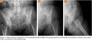

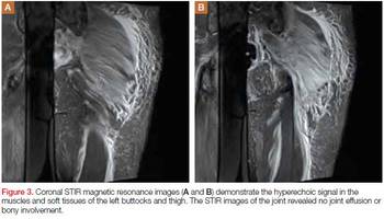
A plain radiograph of the pelvis and left hip showed soft-tissue swelling but no bony pathology or fracture (Figure 1). There was no visual evidence of subcutaneous gas, and chest radiograph was negative for any active disease. Based on the clinical picture of septic shock due to possible septic arthritis, emergent magnetic resonance imaging (MRI) of the left hip and thigh was performed without contrast (Figures 2 and 3). Extensive edema was noted diffusely throughout patient’s left gluteus medius muscle and vastus latera lis muscle; no loculated fluid collection was noted. The edema was concentrated around the proximal left thigh but extended to the level of the knee. The left hip was free of any fluid collection, ruling-out septic arthritis. Based on these MRI findings, a computed tomography scan was ordered, which showed no fluid collection in the pelvis and abdomen (Figure 4).