
Something is Afoot


For each case, choose the correct answer
For answers, see next issue; for discussion, go to www.mdedge.com/clinicianreviews/picture
Case 1 was originally published in the American Journal of Orthopedics (2013;42[3]:121-124).
Case 2 adapted courtesy of Cutis (2000;66[3]:211-212). Copyright 2017 Frontline Medical Communications Inc.
Case 3 adapted from the Journal of Family Practice (2008;57[5]:321-323).
Case 4 credit: ScienceSource. IMAGE NUMBER: SN0826 LICENSE: RIGHTS MANAGED
Answers to last month’s “Picture This” (Clinician Reviews. 2017;27[6]:43): 1c, 2a, 3d, 4b
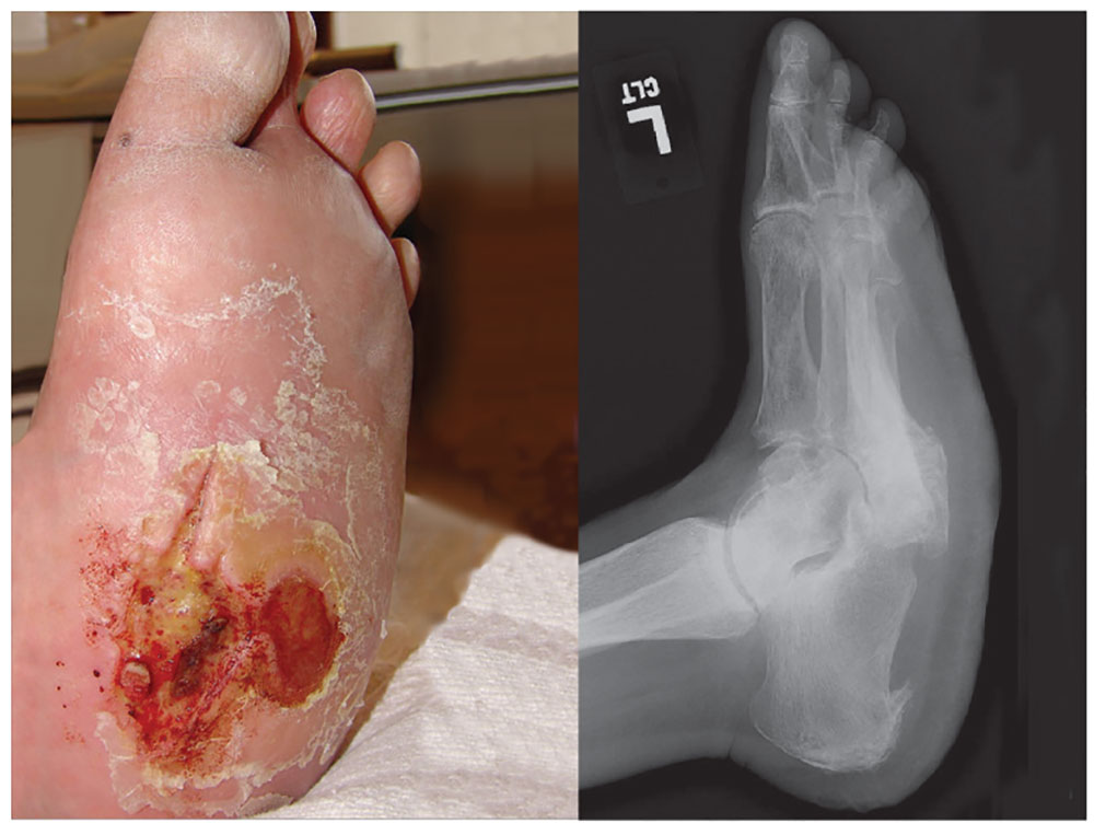
4. A 60-year-old woman seeks relief for a foot wound of several months’ duration that persists despite use of antibiotics and proper care. The skin over the plantar surface has a full-thickness ulcer, with partial necrosis of the subcutaneous tissue. Her history is significant for diabetes with neuropathy, nephropathy, and retinopathy. The diagnosis is
a) Osteomyelitis
b) Deep venous thrombosis
c) Charcot joint
d) Septic joint
Diagnosis: Charcot joint changes, and an associated stage III pressure ulcer, account for the extensive collapse of the inner arch and “rocker bottom foot” seen on the radiograph. Also known as neurogenic arthropathy, Charcot joint is commonly seen with diabetic neuropathy. In affected patients, secondary degenerative changes to the joints occur with loss of normal muscle tone, proprioception, temperature perception, and pain perception. The joints become loose, enlarged, and boggy. There can be extensive cartilage erosion or osteophyte formation. The normal plantar and heel forces are increased, producing eccentric loading of the foot and leading to microfractures, ligament laxity, and bone destruction.
For more information, see “A disfigured foot with ulcer.” J Fam Pract. 2008;57(5):321-323.


