
Noninfectious Penile Lesions


Diagnosis of noninfectious penile cutaneous lesions is guided by the clinical presentation and appearance. In your review, beware the benign lesions that can mimic carcinoma.
Match the diagnosis to the photo by letter
a. Lichen sclerosus
b. Carcinoma in situ
c. Angiokeratoma of Fordyce
d. Seborrheic dermatitis
For answers, see next issue; for discussion, go to www.mdedge.com/clinicianreviews/picture
4. This circumscribed inflammatory plaque on the glans penis of allegedly > 20 years' duration was refractory to circumcision and local treatment. Because of unresponsiveness of the lesion to circumcision and focal steroid infiltration, repeated biopsies were performed.
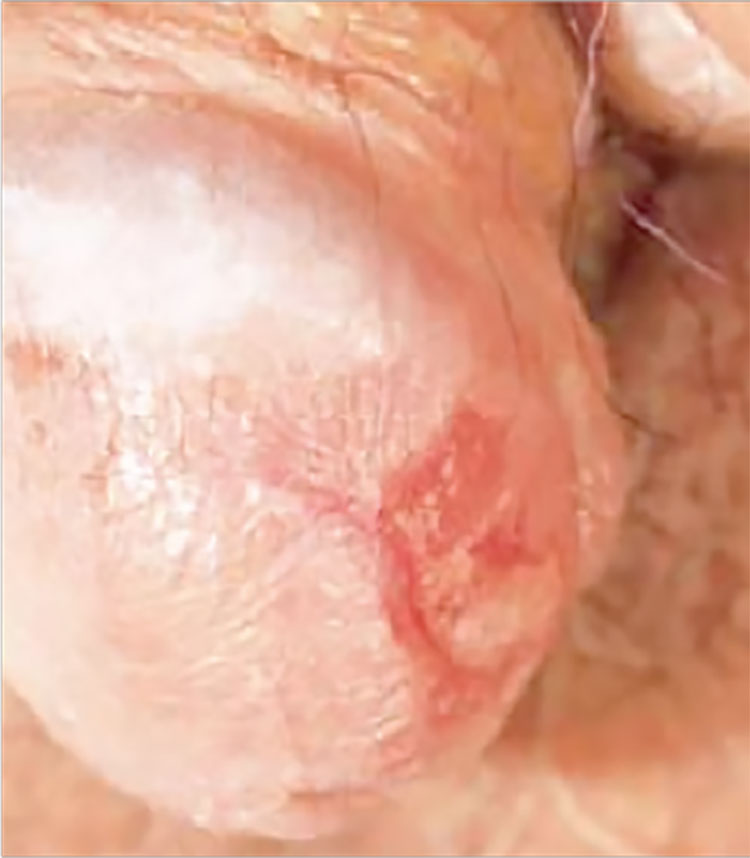
Diagnosis: One biopsy again showed the features of a plasmacellular inflammation, while the other finally revealed the histopathologic features of erythroplasia of Queyrat (carcinoma in situ or Bowen's disease of the glans penis). We assume that either the former biopsy specimens were taken from a plasma cell-rich reactive infiltrate around the neoplastic lesion, or that carcinoma in situ may have arisen due to the chronic inflammation of Zoon's balanitis plasmacellularis. Radiotherapy was performed with good clinical response and subsequent histopathologic proof of complete remission of the lesion.
For more information, see “Bowen's Disease of the Glans Penis (Erythroplasia of Queyrat) in Plasma Cell Balanitis.” Cutis. 2000 June;65(6):395-398.


