
Biomechanical Evaluation of a Novel Suture Augment in Patella Fixation


Patella fractures, although uncommon in the context of corresponding long bone fractures, carry a disproportionately high degree of morbidity, and are often challenging to treat. As such, this study sought to evaluate the efficacy of a Krackow suture augment when compared to standard tension band fixation and cerclage suture augment in patella fracture repair. Cadaveric patella extensor mechanisms were used for biomechanical testing. Specimens were divided among 3 groups, each with a different repair technique: modified anterior tension band (MATB), MATB plus cerclage suture, and MATB plus Krackow suture. Specimens were biomechanically tested in both cyclic and maximum load settings. Mean displacement and load-to-failure forces were measured for cyclic and maximum load testing, respectively. Data was then analyzed with both one-way analysis of variance and independent t-testing. Both augmentation techniques showed improved strength in both cyclic and maximum load testing, with the Krackow suture augment showing the greatest strength. In cyclic testing, cerclage augment showed a 30% decrease in mean displacement while Krackow suture augment showed a 40% decrease when compared to the MATB repair group. Likewise, in maximum load testing, cerclage repair showed a 5% increase and Krackow a 14% increase in load-to-failure force when compared to MATB. Likely due to small sample size, the increases in repair strength did not reach statistical significance. This study provides support for the use of a Krackow suture augment in patella fracture repair, and we suggest this technique may be most useful in the setting of poor bone quality where conventional repair techniques are limited. Although failing to reach statistical significance, these results are encouraging and warrant further investigation in both biomechanical and clinical settings.
Materials and Methods
Specimen Preparation
Fresh-frozen cadaver extensor mechanisms (quadriceps tendon, patella, surrounding retinaculum, patellar tendon) were kept frozen at –4°C until preparation. Fifteen specimens were selected. Mean (SD) age at death was 68 (10) years (range, 51-85 years). One specimen was excluded for a short patella tendon, which precluded adequate attachment for testing. All specimens were free of overt osseous pathology.
After specimens were thawed overnight, the patellae were transversely osteotomized with an osteotome at the junction of the middle and distal thirds of the patella. Sharp dissection was performed to carry the division through the medial and lateral retinaculum at the same level. All 14 specimens were then repaired using the MATB technique. First, the transverse fracture was reduced with a reduction clamp. Then, two 4-mm cannulated screws (DePuy Synthes) were inserted parallel to each other and perpendicular to the fracture. An 18-gauge stainless steel wire was then passed through each screw, crossed anteriorly, and tightened to create a figure-of-8 ATB. The specimens were then randomly divided into 3 groups—MATB; MATB with cerclage suture augment; MATB with Krackow suture augment—while ensuring specimens from a single cadaver were placed in different groups to avoid confounding based on bone density differences.
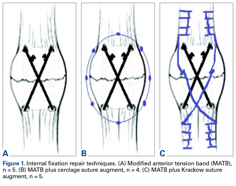
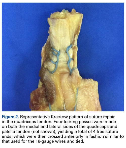
Experimental Setup
Repaired specimens were secured with tissue clamps at the quadriceps and patellar tendons on an MTS Bionix 858 (MTS Systems) hydraulic arm.
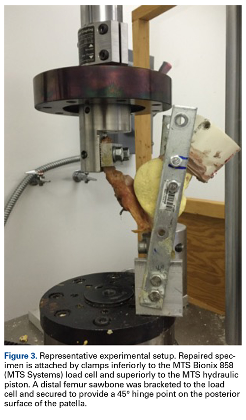
Each patella was secured for cyclic testing. Initially it was placed under 10 N of tension. Then it underwent tensile loading from 10 N to 300 N at 50 N/s for 10 cycles. These parameters were based on previous biomechanical patella studies.10,11 Load was measured with the MTS load cell and displacement with the displacement transducer. Fracture displacement associated with 300-N cyclic tension was recorded. Displacement was calculated as the difference between 10th cycle and 2nd cycle values, which accounted for any degree of initial tissue slippage. After cyclic testing, the patella was placed back in 10 N of tensile loading and subjected to maximum force loading to determine ultimate repair strength. For maximum loading, the patella was stretched progressively at 50 N/s until failure. Again, load and displacement were measured with MTS.
Statistical Analysis
After testing, fracture displacement and maximum load force data were compiled for analysis. One-way analysis of variance with Bonferroni correction was used to determine if there were significant differences between groups. Significance level was set at P < .05.


