
Acral Flat-Topped Papules in an Adolescent Boy


THE DIAGNOSIS: Acrokeratosis Verruciformis of Hopf
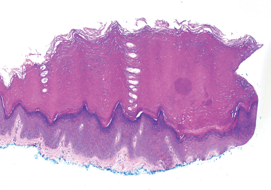
Histopathology from the shave biopsy revealed a circumscribed discrete lesion with papillomatosis, hypergranulosis, and hyperkeratosis. Focal evidence of dyskeratosis was observed in the stratum corneum, and suprabasilar acantholysis also was appreciated (Figure). The patient was referred for genetic testing and was found to have a pathogenic deletion involving exons 1 through 8 of the ATPase sarcoplasmic/endoplasmic reticulum Ca²+ transporting 2 (ATP2A2) gene, leading to a diagnosis of acrokeratosis verruciformis of Hopf (AKV) based on the clinical presentation.
Acrokeratosis verruciformis of Hopf is a rare genodermatosis caused by an autosomal-dominant mutation in the ATP2A2 gene, which directly impacts the sarcoplasmic/endoplasmic reticulum calcium ATPase2 pump and its ability to transport calcium ions.1-3 This ultimately interferes with regulation of epidermal differentiation and intercellular adhesion.3 Acrokeratosis verruciformis of Hopf typically manifests in early childhood, though later onset in adulthood also has been reported.2 The classic presentation of AKV includes multiple small brown to flesh-colored, flat-topped papules arising on the dorsal aspects of the hands and feet. Less commonly, lesions of AKV appear on the arms and legs but do not involve the sebaceous surfaces.2 Additional findings may include nail changes such as longitudinal striations, distal V-shaped nicking, or ridging.2,3
The primary clinical differential diagnosis for AKV includes Darier disease (DD), a genodermatosis with a highly similar pathology to AKV.4 Also referred to as keratosis follicularis, DD carries an autosomaldominant inheritance and arises secondary to mutations in the ATP2A2 gene, affecting sarcoendoplasmic reticulum ATPase type 2 and, consequently, Ca2+ transport, similar to AKV.3,4 Though the relationship between AKV and DD previously has been a source of debate, this common finding supports that AKV and DD are allelic conditions.3 Patients with DD typically develop brown, keratotic papules along seborrheic and intertriginous surfaces within the first 2 decades of life, which may be associated with intense itching, scabbing, and malodor. Patients with DD also may present with nail changes, white mucosal papules, and hyperkeratotic papules on the dorsal hands and feet, representing AKV.4
Additional diagnostic considerations when a pediatric patient presents with flat-topped papules on acral surfaces may include verruca plana, lichen planus (LP), Gottron papules, or epidermodysplasia verruciformis. Verruca plana are most commonly caused by HPV types 3 or 10 and typically arise as flat-topped, flesh-colored to hyperpigmented papules along the extremities, face, and neck. Some patients with verruca plana may have linear arrays of papules due to autoinoculation, which may help to distinguish this condition from AKV.5 Lichen planus represents an inflammatory dermatosis typically manifesting with pruritic flat-topped violaceous papules along the extremities. The presence of Wickham striae may help to distinguish LP from AKV.6 Gottron papules are erythematous lesions that develop over the metacarpophalangeal and interphalangeal joints in the setting of dermatomyositis.7 The presence of additional cutaneous signs of dermatomyositis (eg, the heliotrope sign, shawl sign, holster sign, or nail fold changes) may help to clinically distinguish Gottron papules from AKV. The concentration of papules over the joints of the hands rather than in a more diffuse pattern across the dorsal hands also may help to clinically distinguish Gottron papules from AKV.7 Epidermodysplasia verruciformis is a genodermatosis typically characterized by versicolorlike macules and wartlike papules distributed along the face, neck, trunk, and arms manifesting during childhood.8 The diffuse presentation of lesions, lack of nail involvement and early manifestation of nonmelanoma skin cancers may help distinguish epidermodysplasia verruciformis from AKV.8,9
Dermoscopy can be a useful tool in the initial recognition of AKV. Dermoscopic findings may be particularly useful in challenging cases, such as in patients with skin of color. Characteristic dermoscopic features of AKV include a white reticular pattern containing dotted vessels within the septae as well as areas of white homogeneity.10 This is in contrast to the dermoscopic features of verruca plana (homogenous light-brown to yellow coloration with dotted and/or glomerular vessels), LP (white lines representing Wickham striae, dotted and/or linear vessels, and pink, white, or yellow structureless areas), Gottron papules (dotted vessels, linear branching or curved vessels, patchy white scaling, and white or pink structureless areas), and epidermodysplasia verruciformis (hypopigmented or erythematous background, fine white scaling, dotted vessels, and lightly pigmented vellus hairs).7,10,11
Histopathologic features of AKV include acanthosis, hyperkeratosis, hypergranulosis, and church spire-like papillomatosis, which are considered pathognomonic for AKV.1 While acantholysis (manifesting as suprabasilar clefting) and dyskeratosis (manifesting as corps ronds and grains) have historically been considered histologic features that distinguish DD from AKV, there are reports of acantholysis and dyskeratosis in cases of AKV as well (including in the present case), further supporting a relationship between AKV and DD.1,4 While verruca plana, like AKV, is characterized histologically by acanthosis, hyperkeratosis, and hypergranulosis, verruca plana can be distinguished when koilocytic changes are present. Additionally, verruca plana typically lacks the church spire-like papillomatosis of AKV or verruca vulgaris.5 Lichen planus can be distinguished histologically by the presence of wedgelike hypergranulosis, sawtooth rete ridges, and lichenoid inflammation.6 Histologic features that may help distinguish Gottron papules from AKV include dermal and perivascular lymphocytic infiltrate, basement membrane thickening, mucin deposition, and basal vacuolization.12 EV is notable for mild to moderate epidermal dysplasia, featuring hypergranulosis and hyperkeratosis along with vacuolation and koilocytes.9
Treatment of both AKV and DD is often a temporizing measure as the diseases are chronic in nature without spontaneous remission.3,4 The mainstay of AKV management remains superficial ablation, such as with cryotherapy and laser therapy. Other options may include shave excision, topical retinoids, or systemic retinoids.2 Treatment of DD may include topical therapies such as topical corticosteroids or topical retinoids, systemic retinoids, procedural therapies, and treatment of any secondary cutaneous infections that may develop.4
