Deep brain stimulation: How does it work?


ABSTRACT
Deep brain stimulation has significantly improved the motor symptoms in patients with Parkinson's disease (PD) and other movement disorders. The mechanisms responsible for these improvements continue to be explored. Inhibition at the site of stimulation has been the prevailing explanation for the symptom improvement observed with deep brain stimulation. Research using microelectrode recording during deep brain stimulation in the MPTP monkey model of PD has helped clarify how electrical stimulation of structures within the basal ganglia-thalamocortical circuit improves motor symptoms, and suggests that activation of output and the resultant change in pattern of neuronal activity that permeates throughout the basal ganglia motor circuit is the mechanism responsible for symptom improvement.
THE ‘RATE HYPOTHESIS’: ALTERED CELLULAR DISCHARGE RATES CAUSE PARKINSONIAN MOTOR SYMPTOMS
A good model for PD was lacking prior to the 1980s. As a result, there was little understanding of the pathophysiologic basis for this disorder. A breakthrough in the mid-1980s revolutionized research in this field. A group of young people developed parkinsonian symptoms, and it was discovered that they had all used recreational “designer drugs” containing an impurity: the neurotoxin 1-methyl-4-phenyl-1,2,3,6tetrahydropyridine (MPTP). Now given to primates to simulate PD, MPTP causes all of the classic symptoms of PD except tremor (this may vary from species to species), including freezing, slowness, stiffness, and gait and balance problems. Like humans with PD, primates with MPTP-induced PD even develop dyskinesia after prolonged treatment with levodopa.
Experimentation with MPTP monkeys in the late 1980s led to the “rate hypothesis,” which basically states that when dopamine production is reduced from the substantia nigra compacta (as in PD), changes in striatal activity lead to suppression of GPe activity and a reduction in inhibitory output from the GPe to the STN. This decrease in inhibitory output allows the STN to be overactive, which, in conjunction with a reduction of direct striatal inhibition of the GPi, causes excessive GPi activity and a suppression of thalamic activity to the cortex (Figure 1).
When recording electrodes were placed in these structures in the monkey brain, rate changes were reported to occur in each of these structures in the parkinsonian state.1–4,7 Action potentials recorded from the GPi in MPTP-treated monkeys occurred at a much faster rate than those in healthy monkeys.
Pallidotomy revisited: Dramatic symptom improvement is possible
On the basis of the above and other studies in the MPTP monkey model of PD, investigators in the 1990s reasoned that reduced dopamine in PD led to excessive activity in portions of this circuit. While I would like to say that this led to the rationale for lesioning the STN and GPi for the treatment of PD, this approach had already been taken in the early 1930s and 1940s and continued into the 1960s; it was largely stopped with the introduction of levodopa and was restarted again after the realization that chronic levodopa therapy was associated with a variety of side effects, including the development of excessive involuntary movement and motor fluctuations.
Pallidotomy (lesioning of the pallidum), although tried as a treatment for PD in the 1930s and 1940s, had been abandoned as a result of its inconsistent benefit and lack of effect on parkinsonian tremor. It underwent a resurgence in the 1990s through the work of a group in New York8 that revived Lars Leksell’s pallidotomy approach of the 1960s9 at a time when basic science studies provided the rationale for surgical therapy to create lesions in the GPi. These basic science studies also provided critical new information about the optimal site for lesioning, which led to improved and more consistent outcomes.10–13 In the early years, lesions were created in the anterior (nonmotor) portion of the pallidum but led to inconsistent results. In the 1990s, with a better understanding of the portion of the pallidum involved in motor control, destroying brain tissue by creating a lesion in the posterolateral “motor” region of the pallidum resulted in such dramatic improvement in motor signs that waiting lists of up to 4 years were common for patients who wanted the procedure.
Although unilateral pallidotomy led to marked improvement in motor symptoms on the contralateral side, attempts at bilateral lesions to improve both sides of the body, as well as axial symptoms, were associated with marked hypophonia and, in some reports, cognitive decline. This led physicians and scientists to search for a procedure that could be performed bilaterally without the high incidence of side effects associated with lesioning procedures—and thus to the birth of deep brain stimulation.
Deep brain stimulation as lesion simulation
During the early experience with pallidotomy, the area to be lesioned would first be stimulated with the lesioning probe to observe its effects and thereby determine the precise area in which to create a lesion. At the time, no mechanism existed to leave the stimulator in place rather than create a lesion. But after the development of implantable stimulation devices, chronic stimulation could be delivered bilaterally to the pallidum and STN, resulting in a markedly improved treatment. Since side effects associated with stimulation are reversible, the ability to perform such procedures on both sides of the brain and to adjust stimulation parameters in order to optimize benefits while minimizing side effects made deep brain stimulation the procedure of choice for patients with advanced PD and led to its exploration for treatment of other neurologic disorders.
Because stimulation produced the same or similar benefit as a lesion, most physicians thought that stimulation must work in a similar manner, ie, by decreasing output from the stimulated structure. The rationale for this hypothesis received support from the “rate” model of PD, which postulated that PD motor symptoms occur as a result of overactivity in the STN and GPi. It was postulated that deep brain stimulation improved clinical symptoms by suppressing output from the stimulated structure—in other words, deep brain stimulation effectively caused a physiologic ablation.14,15
FURTHER RESEARCH GIVES RISE TO THE ‘PATTERN HYPOTHESIS’
Deep brain stimulation in the monkey model
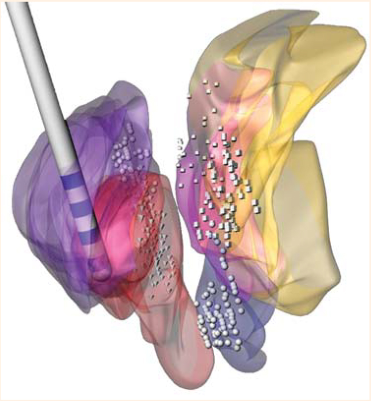
To test the effects of deep brain stimulation, we have performed it in primates with MPTP-induced parkinsonism. Custom-made leads sized to fit a monkey brain are implanted in the same deep brain structures that are targeted when treating PD in humans. Each animal lead has four contacts 0.5 mm in size. We implant a pulse generator, connect the pulse generator to the lead, and set stimulation parameters to improve motor symptoms to mimic a human therapeutic setting as closely as possible. We then record from the basal ganglia structures before, during, and after stimulation that improves the monkey’s motor symptoms. This allows us to determine which changes in neuronal activity in the basal ganglia circuit during stimulation are associated with an improvement in motor symptoms.
In earlier studies examining the mechanism underlying deep brain stimulation, neural activity was recorded only after stimulation, so that activity that occurred during stimulation had to be inferred from that which occurred immediately after stimulation was stopped. We developed a method to subtract artifact produced from stimulation without losing data. This method has been validated, is now used in a number of laboratories, and has revolutionized our ability to study the effect of stimulation on neuronal activity.17
A paradoxical finding
Based on the rate hypothesis, we expected that increased output from the GPi would cause parkinsonian symptoms and predicted that stimulation of the STN should suppress its output, which would suppress excitatory activity to the GPi from the STN and thereby reduce its output. Reduction of the inhibitory output from the GPi to the thalamus would, in turn, lead to a restoration of thalamocortical function and a reduction in the motor signs associated with PD. However, stimulating the STN was found to increase GPi activity.18 Despite increased rates, the incidence and intensity of symptoms were reduced. Further complicating the picture, we were contemporaneously exploring the effect of creating lesions in other parts of the basal ganglia that also led to increased rates of GPi activity, but in this case we observed that the increased rates were associated with a worsening of motor symptoms. In short, we had two laboratories working in parallel that had apparently obtained opposite results: increased GPi activity was associated with improved symptoms in one laboratory and with worse symptoms in the other.18,19