Eosinophilic esophagitis: An increasingly recognized cause of dysphagia, food impaction, and refractory heartburn
ABSTRACTEosinophilic esophagitis is an increasingly recognized cause of a variety of esophageal symptoms, including dysphagia, food impaction, atypical chest pain, and heartburn that does not respond to medical therapy. Its cause is unknown, but allergic and immunemediated mechanisms similar to those of asthma and other atopic diseases are implicated.
KEY POINTS
- The diagnosis is made with upper endoscopy and esophageal biopsies that show diffuse infiltration of eosinophils.
- Current treatment in adults is limited and consists of either swallowed fluticasone (Flonase) or a proton pump inhibitor.
- Because many patients with eosinophilic esophagitis have atopic disease, a complete evaluation for dietary allergens and aeroallergens is recommended, as avoidance of these allergens may be helpful in some adults.
- Cautious endoscopic dilation is a treatment option in patients with evidence of esophageal stenosis. Systemic corticosteroids and novel biologic therapy have been used in refractory cases.
THE ROLE OF ENVIRONMENTAL ALLERGENS AND GENETICS
Studies in children suggest that food allergies are a major contributor to eosinophilic esophagitis. In children, a strict amino-acid elemental diet has led to complete resolution of symptoms and a marked decrease in esophageal eosinophils. However, symptoms tend to recur once patients resume a regular diet.12
It is unclear if dietary modification is effective in adults. In six adults with eosinophilic esophagitis and a history of wheat and rye allergies, symptoms did not improve when these foods were eliminated and did not worsen when they were reintroduced.13
Of interest, there may be a seasonal variation of eosinophilic esophagitis, as suggested by a case report of a 21-year-old woman who had eosinophilic esophagitis that worsened symptomatically and histologically during the pollen season but resolved during winter. This is another example of the role aeroallergens may play in this disease.14
Evidence of a genetic predisposition to this disease is also growing, with a number of case reports describing multiple affected family members spanning generations.15
NEW CONSENSUS ON DIAGNOSTIC CRITERIA
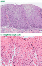
The diagnosis of eosinophilic esophagitis is made histologically, with “marked” eosinophilia on esophageal biopsies, ie, usually 15 or more eosinophils per high-power field. In contrast, a normal esophagus contains almost no eosinophils,16 and esophageal biopsies of patients with GERD usually have fewer than 10 eosinophils per high-power field, with eosinophils limited to the distal esophagus.17
However, a recent systematic review of the literature found 10 different histologic definitions of eosinophilic esophagitis, ranging from more than 5 to more than 30 eosinophils, and more than one-third of the articles included in the review did not contain any specific diagnostic criteria. Similarly, a lack of consensus on the size of a high-power field (ranging from 0.12 to 0.44 mm2) resulted in a 23-fold variability in the description of eosinophil density. Moreover, the biopsy protocols were reported in only 39% of the articles.18
In view of the growing interest in this disease, its increasing recognition, the diagnostic ambiguity described above, and concern about the role of acid reflux, consensus recommendations for its diagnosis and treatment in adults and children have recently been published.19 The current consensus definition for eosinophilic esophagitis is:
- Clinical symptoms of esophageal dysfunction (eg, dysphagia, food impaction);
- At least 15 eosinophils per high-power field; and
- Either no response to a high-dose proton pump inhibitor or normal results on pH monitoring of the distal esophagus.
CLINICAL PRESENTATION
Eosinophilic esophagitis predominantly affects men between the ages of 20 and 40, but cases in women and in younger and older patients have also been reported. Recent systematic reviews found a male-to-female ratio of approximately 3:1.
More than 90% of adults with eosinophilic esophagitis present with intermittent difficulty swallowing solids, while food impaction occurs in more than 60%. Heartburn is the only manifestation in 24% of patients. Noncardiac chest pain, vomiting, and abdominal pain have also been seen, but less frequently.
Up to 80% of patients with eosinophilic esophagitis have a history of atopic disease such as asthma, allergic rhinitis, or allergies to food or medicine. One-third to one-half of patients have peripheral eosinophilia, and up to 55% have increased serum levels of immunoglobulin E (IgE).21
In children, presenting symptoms vary with age and include feeding disorders, vomiting, abdominal pain, and dysphagia. Moreover, children with eosinophilic esophagitis have a higher frequency of atopic symptoms and peripheral eosinophilia than do adults.5,22
Although motor abnormalities are common in patients with eosinophilic esophagitis (up to 40% of patients have esophageal manometric abnormalities, including uncoordinated contractions and ineffective peristalsis),21 esophageal manometry is of limited diagnostic value and so is not recommended as a routine test.19
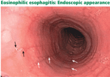
These findings support the theory that inflammation can lead to submucosal fibrosis, remodeling, narrowing, and eventually symptoms. Furthermore, two recent studies found that children with eosinophilic esophagitis had increased subepithelial collagen deposition in their biopsy specimens,24 suggesting increased potential for fibrosis. Also increased are transforming growth factor beta (a profibrotic cytokine) and vascular cell adhesion molecule 1, which is implicated in angiogenesis.25
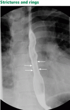
Although many patients with eosinophilic esophagitis have abnormal findings on barium radiography, the test is most useful before esophagogastroduodenoscopy to determine whether a stricture is present and potentially to guide endoscopic dilation.19


