Skin manifestations of diabetes


ABSTRACTDiabetes mellitus can be complicated by a variety of cutaneous manifestations. Good metabolic control may prevent some of these manifestations and may support cure. Unfortunately, most glucose-lowering drugs also have cutaneous side effects. It is important to be able to recognize these signs and symptoms and to either treat them appropriately or refer the patient to a dermatologist or diabetologist.
KEY POINTS
- Patients with type 2 diabetes more often develop skin infections, whereas those with type 1 more often have autoimmune-related lesions.
- Insulin signaling supports normal skin proliferation, differentiation, and maintenance, and a lack of insulin may lead to impaired wound healing, which may affect insulin resorption.
- Skin manifestations of diabetes may also serve as ports of entry for secondary infection.
- A candidal infection (moniliasis) can be an early sign of undiagnosed diabetes.
- Watch for dermal side effects of insulin injections and oral hypoglycemic drugs.
Calciphylaxis
Calciphylaxis is a small-vessel vasculopathy accompanied by mural calcification with intimal proliferation, fibrosis, and thrombosis. It occurs mostly in patients with renal failure and causes a spectrum of end-organ damage due to ischemia. The reported prevalence is 1% to 4% in the dialysis population.
Damage is seen in the epidermal and the subcutaneous tissues. First, redness and tenderness evolve in a small area, which may be surrounded by ecchymosis or pallor, and eventually ischemia leads to the development of subcutaneous nodules and poor-healing, necrotizing skin ulcers. These ulcers serve as a port of entry for infectious agents.
Calciphylaxis has a predilection for vascular regions with thicker subcutaneous adipose tissue, such as the breasts, abdomen, and thighs. In renal failure patients, those who are women, white, obese, or diabetic (especially those with type 2 diabetes) are considered at risk.
Histologic features are medial wall calcification and fibrous expansion in capillaries, venules, arterioles, and small arteries of dermis and subcutaneous fat. Calciphylaxis should not be considered a small-vessel variant of Mönckeberg calcification, which is a medial wall calcification of medium and large vessels. Mönckeberg calcification has been described in patients with diabetes, renal failure, or vitamin D intoxification.
The outcome of calciphylaxis is poor because of impaired wound-healing and infection of the skin with progression to sepsis. Extremely aggressive treatment with analgesics is required for ischemic pain. Furthermore, weight reduction and aggressive control of blood sugar levels seem prudent.35
Eruptive xanthoma
Eruptive xanthoma presents as crops of small (1- to 2-mm) yellow papules with an erythematous halo; these papules may be pruritic and tender. They occur in less than 0.1% of diabetic patients.36 Areas of predilection are extensor surfaces and the buttocks.20
The key histologic feature is the formation of foam cells in the superficial dermis that are mixed with a lymphocytic and neutrophilic infiltrate.
Eruptive xanthomas appear in association with elevated levels of triglyceride-rich lipoproteins. The lipid changes appear in association with familial hypertriglyceridemia and diabetes mellitus, resulting in hypertriglyceridemia from a lack of lipoprotein lipase activity and impaired clearance of chylomicrons and very-low-density lipoproteins. These eruptive xanthomas tend to resolve with control of carbohydrate and lipid metabolism.5,6,17
Granuloma annulare: Not linked to diabetes
Although many have tried to prove an association between localized granuloma annulare and diabetes, no association has been clearly established, and the association between generalized (disseminated) granuloma annulare and diabetes is controversial.21
The cause is not known. The lesions are oval or ring-shaped, with a raised border of skin-colored or erythematous papules. The size varies from millimeters to centimeters. The dorsa of the hands and arms are the areas usually affected. Histologically, the epidermis usually appears normal, whereas the upper and mid dermis show focal degeneration of collagen, palisaded histiocytes around collagen bundles, and abundant mucin.
Localized lesions often resolve spontaneously, whereas the generalized form has a more protracted course which, in rare cases, resolves spontaneously. Sporadic therapeutic success has been reported with topical, systemic, and intralesional steroids; isotretinoin; chlorambucil (Leukeran); cryotherapy; chlorpropamide (Diabinese); chloroquine; potassium iodide/nicotinamide; dapsone; antimalarials; and psoralen-ultraviolet A light.5,6,20
CUTANEOUS INFECTIONS
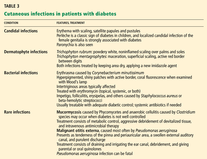
Candidal infection
A candidal infection (moniliasis) can be an early sign of undiagnosed diabetes. Perlèche is a classic sign of diabetes in children, and localized candidal infection of the female genitalia has a strong association with diabetes. This infection appears as erythema with scaling and typical satellite papules and pustules. Paronychia is another sign.
It is important to remember that in men, Candida balanitis, balanoposthitis, and intertrigo can be presenting signs of diabetes.
Candidal infections improve with adequate metabolic control and treatment with topical midazoles or nystatin (Mycostatin).5,37
Infections with dermatophytes
Common superficial infections are caused by Trichophyton rubrum, T mentagrophytes, and Epidermophyton floccosum. In diabetic patients, onychomycosis or tinea pedis needs to be monitored for and treated, as it can be a port of entry for infection. This is especially true for patients with neurovascular complications and intertrigo.
Signs of T rubrum infection are noninflamed, white, powdery scaling or skin creases on the palms and soles, often with nail involvement. T mentagrophytes-associated intertrigo or interdigital infection presents as maceration and superficial scaling with an active red border. Treatments of choice are drying the local area and applying one of the newer topical imidazole antifungal agents.5,37