Congenital uterine anomalies: A resource of diagnostic images, Part 1


Accurate diagnosis of such anomalies has prognostic importance for both obstetric and gynecologic outcomes
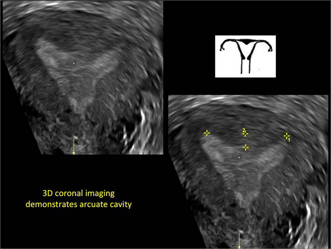
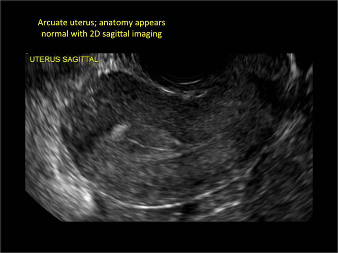
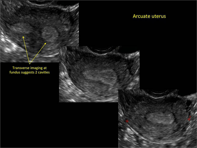
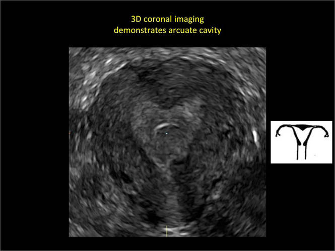
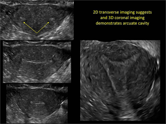
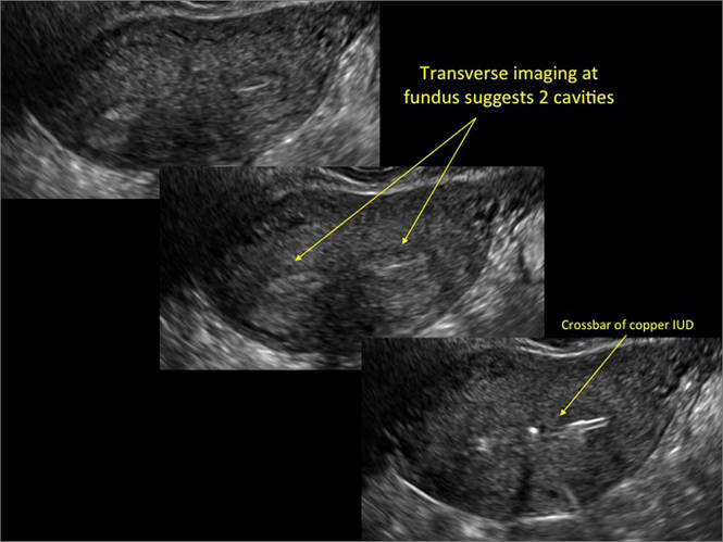
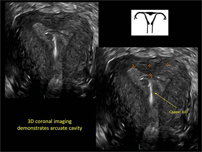
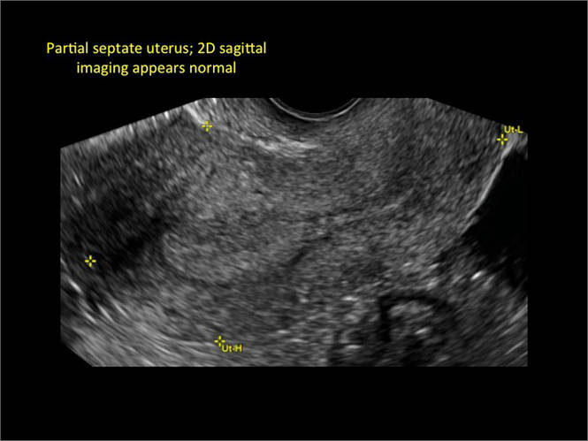
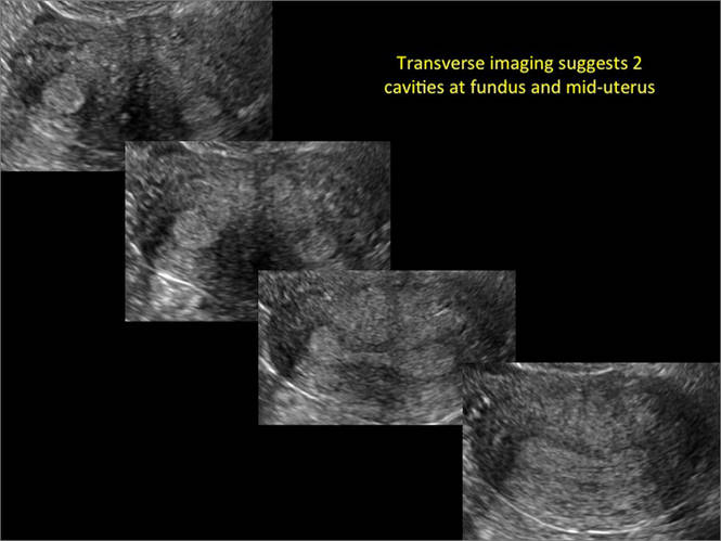
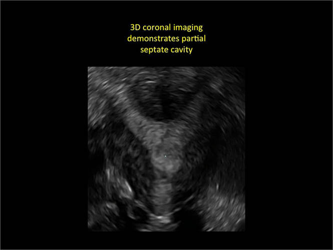
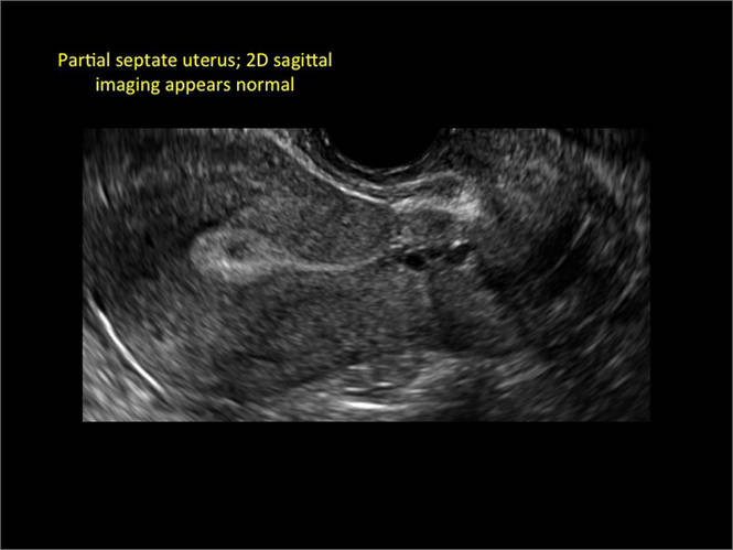
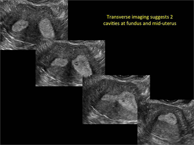
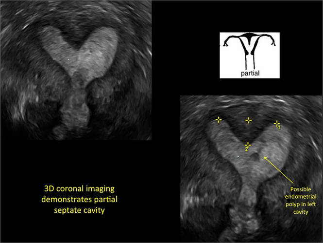
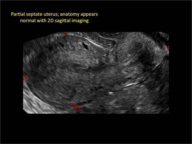
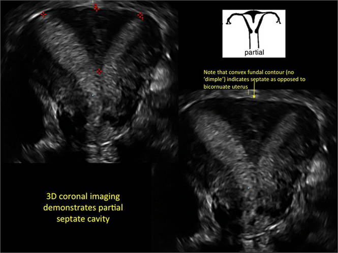
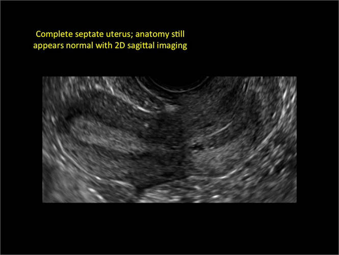
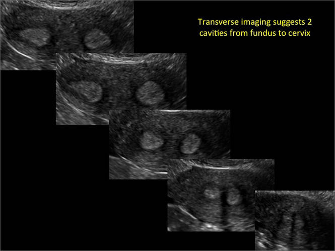
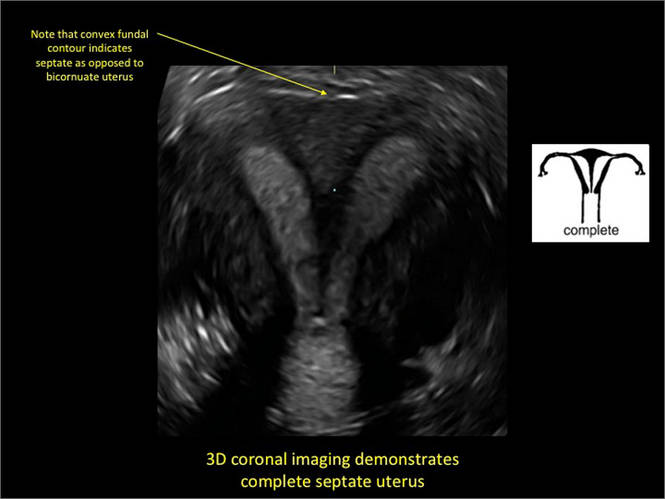
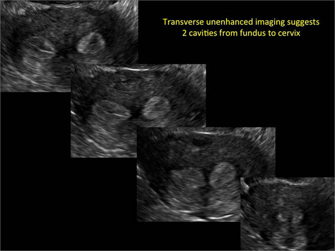
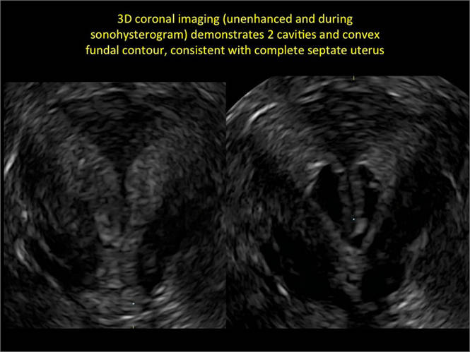
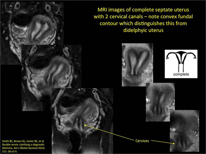
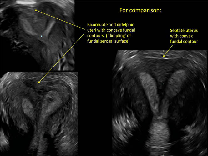
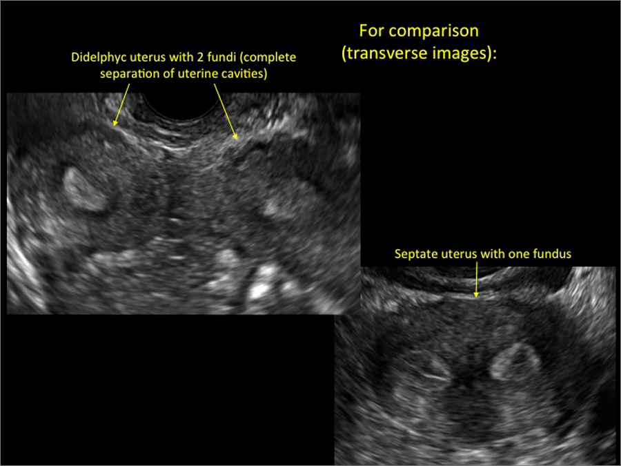
Case: Partial septate uterus
|
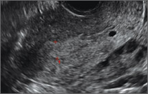
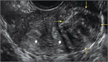
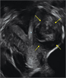
ADDITIONAL IMAGES


Share your thoughts on this article! Send your Letter to the Editor to rbarbieri@frontlinemedcom.com. Please include your name and the city and state in which you practice.