Drew C. Baird, MD Department of Family and Community Medicine, D.D. Eisenhower Army Medical Center, Fort Gordon, Ga drew.baird@us.army.mil
EDITOR Richard P. Usatine, MD University of Texas Health Science Center at San Antonio
The author reported no potential conflict of interest relevant to this article. The opinions or assertions contained herein are the private views of the author and are not to be construed as official or as reflecting the views of the Department of Defense.
This patient’s leg pain was the tip of the iceberg. He also had diminished breath sounds and a cachectic appearance.
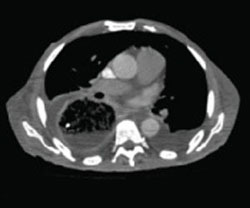
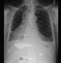
In our patient’s initial workup, a CT (FIGURE 2), barium study (FIGURE 3), and endoscopy were all used to rule out an obstructive lesion.
FIGURE 2 Massively distended esophagus
A chest CT revealed a distended esophagus—at times 8 cm in diameter—from the thoracic inlet to the gastric inlet, strongly suggestive of achalasia. The patient’s trachea was displaced anteriorly, his posterior right lung was compressed, he had bilateral pleural effusions, and had generalized edema that was suggestive of anasarca.
FIGURE 3 The “bird’s beak” of achalasia
A barium swallow study showed diffuse enlargement of the esophagus with a narrowing “bird’s beak” near the esophagogastric junction, consistent with achalasia.
Treatment targets the constricted LES
The goal of treatment in achalasia is to help food move through the constricted LES. Pharmacologic therapy, used to decrease LES resting tone, has limited benefit and becomes less effective as the disease progresses. Calcium channel blockers, particularly nifedipine, and nitrates are the most commonly used agents.5