Tumor Volume: An Adjunct Prognostic Factor in Cutaneous Melanoma


Measurement of tumor volume may be a helpful adjunct to established prognostic factors in cutaneous melanoma, including Breslow depth, presence or absence of ulceration, mitotic index, lymphovascular invasion, and microsatellites. This report expands on the theory that a tumor volume cutoff point of 250 mm3 as measured by surface area of the lesion (ie, longest vertical and horizontal measurements either based on clinical or gross pathological assessment) multiplied by the Breslow depth could serve as a potentially relevant predictor of sentinel lymph node (SLN) metastasis in both thin and thick invasive cutaneous melanomas, which prompted investigation of a larger sample size using the pathology database at our institution.
Practice Points
- Measurement of melanoma tumor volume using clinical area (length • width of the lesion before diagnostic biopsy) multiplied by Breslow depth may provide additional prognostic information.
- Further study is needed to validate the use of tumor volume as an adjunct to established histopathologic prognostic factors in cutaneous melanoma.
Results
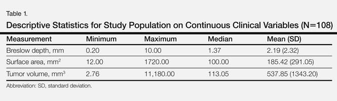
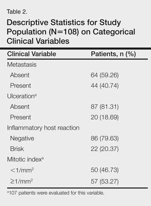
There were 108 eligible cases in the 4-year time period in which tumor volume assessment could be determined based on the pathology report in conjunction with Breslow depth, mitotic index, presence or absence of ulceration, and tumor infiltrating response. Breslow depth ranged from 0.20 to 10.00 mm, with a median depth of 1.37 mm. Surface area ranged from 12.00 to 1720.00 mm2 (median, 100.00 mm2). Tumor volume was calculated by multiplying Breslow depth by surface area and ranged from 2.76 to 11,180.00 mm3 (median, 113.05 mm3)(Table 1). Ulceration was present in 18.69% of the tumors, 20.37% exhibited a brisk inflammatory host reaction, and 53.27% had a mitotic index of 1/mm2 or more. Tumor metastasis was noted in 40.74% (44/108) of patients (Table 2), all of whom had a primary melanoma with a Breslow depth greater than 1 mm. Only one T1 melanoma had a tumor volume greater than 250 mm3. Metastasis in patients with T2 (1- to 2-mm thick) and T3 (2- to 4-mm thick) melanoma was associated with a tumor volume greater than 250 mm3 in 16 of 26 patients (61.54%), and all 18 patients with T4 melanomas (>4-mm thick) had tumor volume greater than 250 mm3.