
When Skin Cancer Skips the Skin: Merkel Cell Carcinoma Presenting With Isolated Lymphadenopathy


Background: Merkel cell carcinoma (MCC) is a rare, aggressive cutaneous neuroendocrine tumor with a high mortality rate and increasing incidence, particularly in older, immunosuppressed, and fair-skinned adults. Though most cases present with a primary skin lesion, MCC can rarely manifest solely as nodal metastases without an identifiable dermatologic origin, a presentation known as MCC of unknown primary lesion.
Case Presentation: A 70-year-old male with multiple comorbidities presented with progressive right-sided jaw swelling and dysphagia. Physical examination revealed localized edema and a firm, mildly tender submandibular mass without an evident primary skin lesion. Initial management for presumed parotitis failed to resolve symptoms. Imaging revealed necrotic submandibular and cervical lymphadenopathy, and subsequent biopsy confirmed a high-grade neuroendocrine carcinoma consistent with MCC.
Conclusions: This case underscores the importance of including MCC in the differential diagnosis of rapidly enlarging cervical masses, particularly in older adults, even in the absence of a cutaneous lesion. Early biopsy and immunohistochemical analysis are critical for accurate diagnosis. This case also highlights the clinical challenge of managing MCC in patients with comorbidities and limited treatment options, emphasizing the value of a multidisciplinary approach.
Merkel cell carcinoma (MCC) is a rare but highly aggressive primary cutaneous neuroendocrine carcinoma with a poor prognosis and a 2-year mortality rate of 28%, surpassing melanoma. Due to its rapidly progressive nature and potential for early metastasis, prompt recognition and intervention are essential.1 Incidence rates of MCC have been increasing significantly, with about 2488 cases reported in the United States in 2013, a 95% increase from 2000. Demographic projections estimate > 3000 new US cases in 2025. MCC primarily affects older adults with a mean age of 75 years, and its incidence increases exponentially with age. Light skin is a strong risk factor, with most cases occurring in White individuals.1-3
MCC is derived from Merkel cells, mechanoreceptors located in the basal epidermal layer of the skin. MCC pathogenesis is linked to Merkel cell polyomavirus (MCPyV) and UV radiation-induced mutations. MCPyV integrates into the genome, driving uncontrolled cell growth, while UV exposure leads to high mutational burden in virus-negative cases. Immunosuppression is another key factor; patients with compromised immune function tend to develop MCC at a younger age and experience worse outcomes.4,5
Clinically, MCC presents as a rapidly enlarging, violaceous, and painless nodule on sun-exposed areas, predominantly in older adults. Due to its nonspecific appearance, early diagnosis is challenging. MCC of unknown primary lesion (MCC-UP) is a rare subtype characterized by the absence of an identifiable primary cutaneous tumor, with cancer instead presenting in lymph nodes or other metastatic sites. Histologically, MCC is composed of small, round blue cells with scant cytoplasm, finely granular chromatin, and small nucleoli, typically arranged in sheets or trabeculae. Immunohistochemical (IHC) markers such as synaptophysin, chromogranin, CD56, and cytokeratin 20 (CK20) help confirm the diagnosis.6
MCC staging is based on tumor size and metastasis. Sentinel lymph node biopsy is a key prognostic tool that significantly impacts disease management.7 Treatment for MCC typically involves wide local excision with or without adjuvant radiotherapy for localized disease. In advanced cases, immune checkpoint inhibitors targeting the programmed cell death 1/programmed cell death ligand 1 pathway have shown promising results in improving survival. Given the aggressive nature of MCC, early detection and a multidisciplinary approach are crucial.8
This report presents an aggressive case of MCC without an identifiable primary cutaneous lesion involving the submandibular and cervical lymph node chains. Additionally, we provide a brief literature review to better contextualize this rare disease manifestation.
Case Presentation
A 70-year-old male presented to a skilled nursing facility with difficulty swallowing, swelling of the right side of his face, and tenderness of the right side of his neck. The patient had a history of atrial fibrillation, hypertension, type 2 diabetes mellitus, deep vein thrombosis treated with apixaban, bipolar disorder, oxygen-dependent chronic obstructive pulmonary disease, and multiple decubitus ulcers. Physical examination revealed localized edema below the right ear and along the right jawline. On palpation, the area was firm and mildly tender to touch, without fluctuance or overlying skin changes.
No primary dermatological lesion was discovered on inspection of the affected region or surrounding skin. Laboratory tests at that time were remarkable for macrocytic anemia (hemoglobin level, 11.7 g/dL; mean corpuscular volume, 103.9 fL) without leukocytosis. The patient declined to undergo imaging. A presumptive diagnosis of parotitis was made and the patient was treated with an empiric course of doxycycline 100 mg twice daily for 10 days. Despite the treatment, his symptoms persisted and the Infectious Disease service extended the antibiotic regimen to 14 days while adding a 7-day course of augmentin 875 mg twice daily.
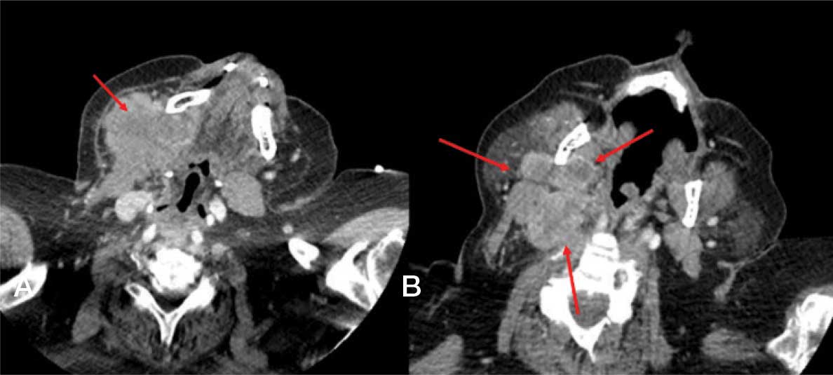
Despite empiric treatment for parotitis, the patient reported worsening symptoms. A physical examination revealed progression of the swelling with increased tenderness to palpation. No oropharyngeal exudate, rashes, or erythema were appreciated. The patient ultimately agreed to a computed tomography of the neck with contrast, which demonstrated large necrotic lymphadenopathy involving the right submandibular and right level IIa stations with largest conglomerates measuring 6.0 × 6.6 cm and 5.2 × 3.3 cm, respectively. There was adjacent narrowing of the internal jugular vein, highly suspicious for malignancy (Figure 1).
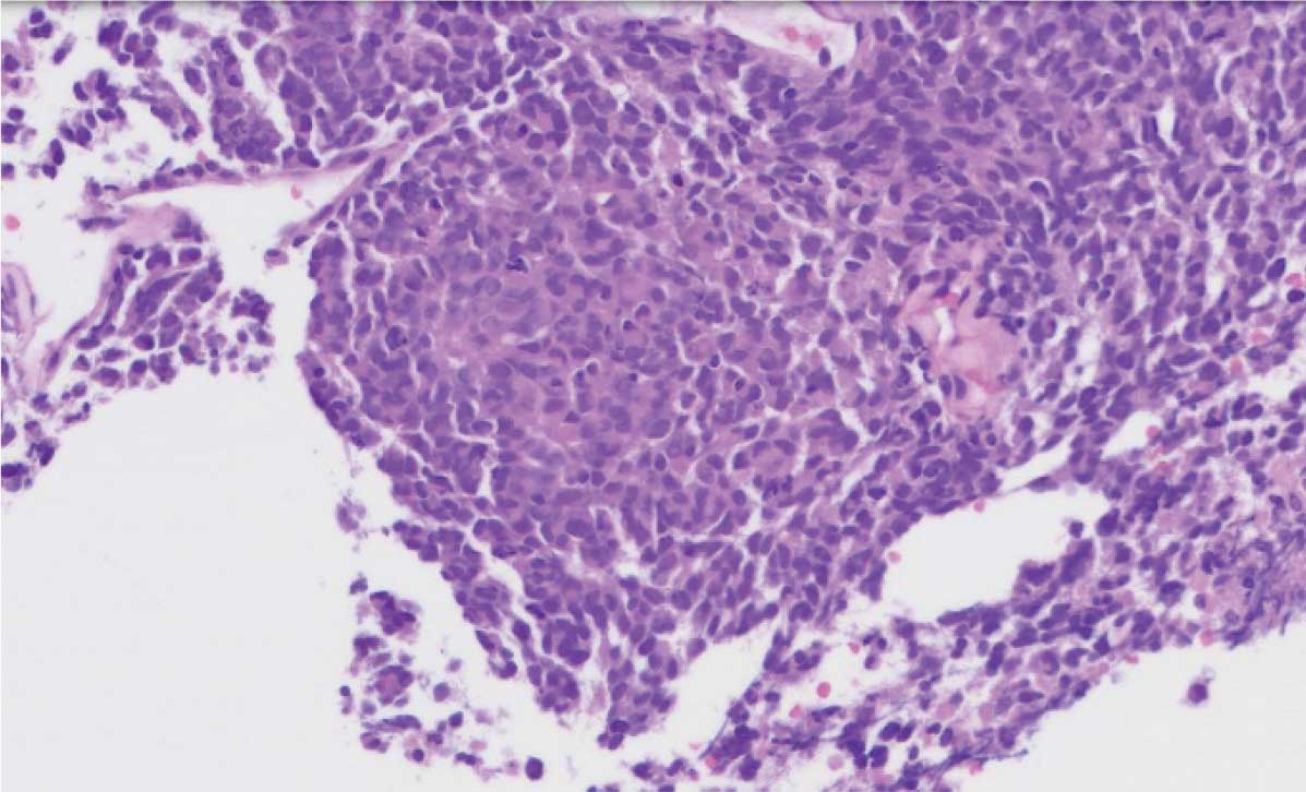
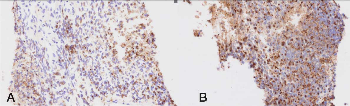
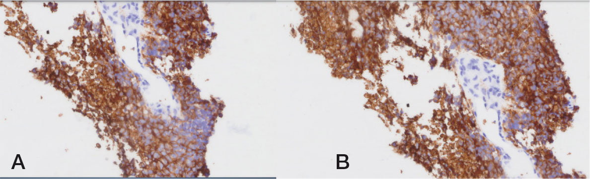
A fine-needle biopsy was performed. Flow cytometry demonstrated CD56+ large cells of probable nonhematolymphoid origin and neuroendocrine differentiation. Cytopathological studies revealed metastatic high-grade neuroendocrine carcinoma, most consistent with MCC (Figure 2). IHC staining confirmed MCC diagnosis, with cells staining positive for pancytokeratin (also referred to as multicytokeratin) with a perinuclear dot-like pattern, CK20 (Figure 3), CD56, and synaptophysin (Figure 4). The cells were negative for CD45, SOX10, S100, TTF-1C, CD138, and CK7, and demonstrated a high Ki-67 proliferative index. A subsequent positron emission tomography revealed a hypermetabolic mass in the right parotid gland and an 89 mm complex mass in the right submandibular space, with enlarged lymph nodes up to the right lower paratracheal node, and a lesion in the left kidney that was equivocal for a solid lesion.
Multidisciplinary discussions included medical oncology and otolaryngology. The patient was determined not to be a candidate for systemic therapy including immunotherapy and surgical resection given his comorbidities and poor performance status (Eastern Cooperative Oncology Group performance status score of 4 and American Society of Anesthesiologists physical status classification IV). The patient was transitioned to hospice care, and although radiation therapy was considered, he died before evaluation could be completed.
Discussion
MCC is a rare, aggressive cutaneous neuroendocrine malignancy. Incidence has increased globally, attributed to an aging population, enhanced diagnostic capabilities, and rising immunosuppressed populations due to conditions such as HIV/AIDS or organ transplantation.2,9 MCC incidence is estimated at about 2500 new cases per year in the US.9 MCC primarily affects older adults and males. The peak incidence is in individuals aged > 65 years. A strong association with UV radiation exposure is noted, and fair-skinned individuals are disproportionately affected. Additionally, infection with MCPyV is implicated in about 80% of cases, highlighting a viral oncogenesis component in the etiology.9,10
MCC derives from cutaneous mechanoreceptor Merkel cells in the basal layer of the epidermis and its pathophysiology involves viral and nonviral mechanisms. MCPyV-associated MCC arises from integration of the virus into the host genome, leading to expression of viral T antigens that drive tumorigenesis.10
UV-induced MCC is characterized by high mutational burdens typical of UV damage, including mutations in tumor suppressor genes such as TP53 and RB1. Both pathways converge on dysregulation of the cell cycle, apoptosis evasion, and uncontrolled proliferation.11 The neuroendocrine origin from Merkel cells is supported by the expression of markers CK20, CD56, and synaptophysin.12 The patient was positive for all 3 neuroendocrine markers, in addition to pancytokeratin. This positive staining, as well as other negative markers for hematologic and melanocytic malignancies underscores the importance of tissue biopsy and IHC in establishing a definitive diagnosis and excluding other differential diagnoses such as metastatic small cell lung carcinoma, lymphoma, and small cell melanoma. Additional diagnostic tools, such as the AMERK serologic test for MCPyV antibodies, are often used in surveillance and prognostication, but were not performed in this case. Since the patient was not a candidate for treatment, MCPyV testing would not have altered clinical management.13
MCC typically presents as a painless, rapidly growing, firm nodule or plaque on sun-exposed areas, particularly the head and neck. In this patient it presented with submandibular and cervical lymphadenopathy, which drain these locations. Lesions are often flesh-colored, violaceous, or erythematous and may ulcerate.12 AEIOU (Asymptomatic, Expanding rapidly, Immunosuppression, Older age [> 55 years], and UV-exposed site on fair skin) highlights key clinical features suggestive of MCC.14 Regional lymph node involvement is common at presentation, with 26% of MCC cases presenting with lymph node involvement and 8% of cases presenting with distant metastases, underscoring the tumor’s aggressive nature.15 The tumor has a 5-year overall survival rate of 17.2% with distant metastasis.16 The patient died 2 months after presentation; however, he had significant comorbidities which may have contributed to his rapid decline.
MCC-UP
MCC-UP is a subtype of MCC characterized by the absence of an identifiable primary cutaneous tumor, with cancer presenting in lymph nodes or other metastatic sites. The patient’s presentation is consistent with MCC-UP. MCC-UP accounts for about 8% to 19% of all MCC cases and poses distinct diagnostic and therapeutic challenges because it often presents at more advanced stages due to the absence of an identifiable primary lesion.17,18
MCC-UP is often associated with a more robust immune response that is thought to facilitate the regression or immune clearance of the primary skin tumor. Notably, the patient had extensive comorbidities (eg, chronic infections such as osteomyelitis), which may have contributed to immune dysregulation and represent an alternative factor influencing this phenomenon.17 By definition, MCC-UP is more likely to present at a more advanced stage.18 Although regional lymph nodes are the most common sites of metastasis in MCC, involvement of other locations—such as the parotid gland—has also been reported.19
Histologically, MCC demonstrates small round blue cells with scant cytoplasm and finely dispersed chromatin, arranged in sheets, nests, or trabeculae. Tumors exhibit high mitotic activity and frequent necrosis.20 Additional features include nuclear molding that occurs when adjacent tumor nuclei indent against one another, and a lack of prominent nucleoli. Differential diagnosis includes other small round blue cell tumors such as small cell lung cancer, necessitating a comprehensive IHC panel to confirm the diagnosis. IHC hallmark markers include CK20 in a dot-like perinuclear pattern, synaptophysin, and CD56.20 MCPyV-positive tumors also demonstrate expression of viral T antigen.10,11 Ki-67, a marker of cellular proliferation, is typically elevated in MCC and reflects its aggressive biological behavior. In the patient, Ki-67 showed a high proliferative index.20
Treatment
Management of MCC involves a multidisciplinary approach tailored to the tumor stage. Surgery with wide local excision and sentinel lymph node biopsy is the cornerstone of therapy for localized disease.2 Adjuvant radiation therapy is commonly employed to reduce recurrence risk, particularly in cases with positive margins or lymph node involvement.14 MCC-UP responds very well to radiation, and systemic therapies are critical for advanced or metastatic disease.18,21 Particularly with MCPyV-associated MCC, immune checkpoint inhibitors, such as avelumab and pembrolizumab, are effective in leveraging immune-mediated tumor control, as these tumors exhibit heightened immunogenicity due to viral antigens.21,22 Traditional chemotherapy remains an option but is associated with limited efficacy and significant toxicity.23
In this case, treatment options were limited; the patient was not a candidate for surgery or systemic immunotherapy due to multiple comorbidities, poor performance status, and rapid clinical decline, and he was unable to undergo radiation therapy. Palliative care and hospice are the preferred approach to management in those cases.
Conclusions
MCC is an aggressive malignancy with increasing incidence in the aging population. Although it typically presents as a cutaneous lesion on sun-exposed skin, a subset of cases classified as MCC-UP presents solely with nodal involvement, complicating timely diagnosis. This report highlights the need for heightened clinical suspicion of MCC in older adults with atypical or treatment-refractory cervical masses. Early tissue diagnosis and staging are essential to allow for the timely initiation of therapy. This case highlights the clinical challenge of managing MCC in patients with significant comorbidities and limited treatment options, underscoring the value of a multidisciplinary approach.
