Authors’ Disclosure Statement: The authors report no actual or potential conflict of interest in relation to this article.
Dr. Houdek and Dr. Sems are Assistant Professors of Orthopedics, Dr. Wagner is an Upper Extremity Fellow, Dr. Watts is an Orthopedic Surgery Resident; and Dr. Moran is a Professor of Plastic Surgery and Orthopedics, Department of Orthopedic Surgery, Mayo Clinic, Rochester, Minnesota. Dr. Houdek was a resident at the time the article was written.
Address correspondence to: Steven L. Moran, MD, Mayo Clinic, Division of Plastic and Reconstructive Surgery, 200 First St. SW, Rochester, MN 55905 (email, moran.steven@mayo.edu).
Am J Orthop. 2018;47(6). Copyright Frontline Medical Communications Inc. 2018. All rights reserved.
Matthew T. Houdek, MD Eric R. Wagner, MD Chad D. Watts, MD Stephen A. Sems, MD Steven L. Moran, MD . Free Composite Serratus Anterior-Latissimus-Rib Flaps for Acute One-Stage Reconstruction of Gustilo IIIB Tibia Fractures. Am J Orthop.
June 20, 2018
TAKE-HOME POINTS
Gustilo IIIB injuries with segmental bone loss can be difficult to treat with conventional means.
Vascularized bone grafts are beneficial for reconstructing bone defects >5 cm.
The SALR composite flap consists of bone and muscle.
This flap can provide soft tissue coverage and vascularized bone in a single surgical procedure.
In this study, the use of the SALR composite flap was capable of healing large segmental bony defects at an average of 7 months.
SURGICAL TECHNIQUE
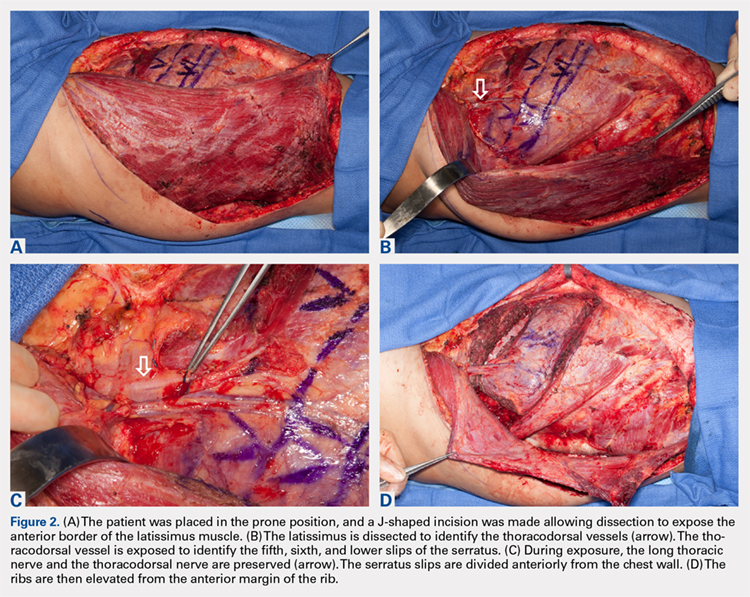
The patient is placed in the lateral decubitus position during the procedure. A 2-team approach is used for dissection of the flap and preparation of recipient vessels to decrease operative time. A J-shaped incision is started on the chest at the mid-axillary line and extended just over the fifth and sixth rib. The incision can be made to fall into the intermammary crease in a woman to hide the scar. The dissection begins by exposing the anterior border of the latissimus muscle (Figure 2A). Next, the latissimus is dissected to reveal the thoracodorsal vessels (Figure 2B). At this level, the thoracodorsal vessel can be traced into the axilla. The branch going into the fifth, sixth, and lower slips of the serratus are dissected. The long thoracic nerve and the thoracodorsal nerve are preserved during the dissection (Figure 2C). The fifth, sixth, and seventh slips of the serratus are preferentially included in the dissection while leaving the most superior slips of the serratus to preserve scapular stability. Dissection begins by identifying 2 adjacent rib sections of the fifth and sixth or sixth and seventh ribs. The defect in the lower extremity determines the length of rib harvested. The serratus slips are then divided anteriorly over the chest wall. The dissection is extended to the intercostal spaces of the fourth and fifth ribs. The supraperiosteal dissection is performed at the anterior margin of the rib (Figure 2D).
The intercostal vessels to the fourth rib are dissected free to preserve vascularity and sensation to the nipple areola complex. The ribs are dissected back at the level of the fifth rib superiorly and the distal aspect of the sixth rib, while being careful to preserve the intercostal vessels with the sixth rib to preserve the periosteal blood supply. The intercostal vessels are ligated as the length of rib is dissected posteriorly. Here the serratus muscle is left intact with the periosteum and now is divided posteriorly. Dissection is then performed around the serratus branch of the thoracodorsal vessels and these are dissected back to the main thoracodorsal trunk (Figure 3A). The small branches of the long thoracic nerve are preserved proximally to maintain the innervation to more proximal segments of the serratus. The serratus is then lifted off the chest wall with the underlying rib and a segment of the intercostal muscle (Figure 3B). The defect in the chest wall can then be reconstructed with synthetic mesh or decellularized dermis (Figure 3C).
The parietal pleura is not violated in most dissections; however, if it is, a temporary chest tube or red rubber catheter can be placed within the thorax and allowed to water seal for 24 hours. The flap may be taken with a portion or all of the latissimus if there is a requirement for significant soft tissue reconstruction. The latissimus, serratus, and rib are all maintained on the thoracodorsal vessels, which are dissected to the level of the axillae. The thoracodorsal vessels are divided once adequate length has been achieved and the flap is transferred down to the lower extremity where the anastomosis is performed preferentially in an end-to-side fashion to the anterior or posterior tibial vessels. Following the anastomosis, the ribs are secured to the defect in an onlay fashion using compression screws. A washer is used in addition to the screw to allow for compression of the graft against the defect. A small partial greenstick fracture can be created over the curved aspect of the convex surface of the rib to enable the rib to straighten more effectively (Figure 4A). Small wedges can be removed from the rib to create a straight bone segment if this is necessary because of the length of the defect. Finally, the donor site in the chest is closed in a layered fashion. The latissimus and serratus portions of the flap are then covered with a split-thickness skin graft once they are secured in the lower leg.