
Four Fattened Phalanges


Match the diagnosis to the photo by letter
a. Digital fibromyxoma
b. Dermatofibrosarcoma protuberans
c. Subungual exostosis
d. Onychomatricoma
For answers, see next issue; for discussion, go to www.mdedge.com/clinicianreviews/picture
Case presentations and photographs courtesy of Cutis. Copyright 2018 Frontline Medical Communications Inc.
Answers to November “Picture This” (Clinician Reviews. 2017;27[11]:39): 1c, 2a, 3b
3. For the past year, a 30-year-old man’s left great toe has had a 3-cm exophytic, yellowish red, subungual nodule that is obliterating the nail plate. Ten years ago, a similar nodule in the same location was removed via laser by a podiatrist. Plain radiographs demonstrate an inferior cortical lucency of the distal phalanx, as well as a lucency over the nail bed with calcification extending to the soft tissues. MRI reveals bone erosion from the overlying mass.
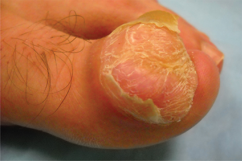
Diagnosis: Digital fibromyxoma is the term used to describe a distinctive, slow-growing, soft-tissue tumor with a predilection for the periungual or subungual regions of the fingers and toes. This benign growth typically presents as a painless or tender nodule in middle-aged adults, with a slight male predominance. In a case series of 124 patients, 36% who had imaging studies showed bone involvement by an erosive or lytic lesion. Soft-tissue invasion of the bone is demonstrated by scalloping on plain radiographs.
,For more information, see “Toe Nodule Obliterating the Nail Bed.” Cutis. 2016;97(4):260, 281-282.


