Clinical features and diagnosis of small-vessel vasculitis


ABSTRACTVasculitis is inflammation of the blood vessel. Granulomatosis with polyangiitis (GPA), microscopic polyangiitis, and eosinophilic GPA are three small-vessel vasculitic diseases that share certain features, but also have important differences. Distinguishing these entities may influence the diagnostic approach, treatment decisions, and outcomes. Circulating antineutrophil cytoplasmic antibodies (ANCA) characterize all three diseases, although their immunofluorescence patterns and target antigen specificities differ. While the presence of ANCA can suggest these diagnoses, the diseases are best viewed as separate entities, each defined by specific clinical and histologic characteristics.
DIFFERENTIATION
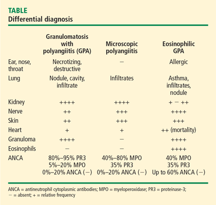
A key histologic difference between GPA and MPA is the presence of granulomatous inflammation in GPA and its absence in MPA under the current nomenclature system.9 Granulomatous inflammation can be seen in EGPA, but it is usually accompanied by eosinophils, which are less likely to be present in GPA and MPA.
The predominance of the ANCA immunofluorescence pattern and target antigen differs between GPA and MPA, with ANCA positivity occurring in 38% of patients with EGPA.13
SUMMARY
Conceptualizing vasculitic disease based on vessel size can be useful, but it is not an absolute definition. Although GPA, MPA, and EGPA predominantly affect small- to medium-sized vessels, these disease entities are phenotypically unique, with both shared features and differences. Common to all three entities is the potential for organ- and life-threatening manifestations, particularly involving the lungs, kidneys, nerves, gastrointestinal tract, and heart. All three entities need aggressive immuno suppression for severe disease. Recognition of these entities and the distinctions among them can guide the approach to diagnosis, treatment, and future outcomes.