ADVERTISEMENT

Difficulty swallowing solid foods; food ‘getting stuck in the chest’

Cleveland Clinic Journal of Medicine. 2010 June;77(6):354-363 | 10.3949/ccjm.77a.09144
Author and Disclosure Information

CASE CONTINUES: HER CONDITION IMPROVES, THEN WORSENS

Figure 2. After treatment with a proton pump inhibitor, clarithromycin, and amoxicillin for 14 days, no nodules are visible.
The patient receives a proton pump inhibitor, clarithromycin, and amoxicillin for 14 days. Her dysphagia improves, and endoscopy with biopsies 3 months later is negative for MALT lymphoma and H pylori (Figure 2).
Figure 3. Follow-up endoscopy demonstrates stomach mucosa with multiple nodules (arrow) due to recurrence of mucosa-associated lymphoid tissue (MALT) lymphoma after treatment for Helicobacter pylori and remission.
However, when she undergoes endoscopy with endoscopic ultrasonography again 6 months after her second endoscopy, it shows multiple 3-to-5-mm smooth nodules in the body of the stomach (Figure 3). Multiple biopsies are then performed; the findings in the gastric body are consistent with extranodal marginal zone B-cell lymphoma of MALT type, t(11;18)-negative. Giemsa stain is negative for H pylori.

Computed tomography of the chest, abdomen, and pelvis reveals no evidence of additional sites of tumor. Positron emission tomography reveals increased uptake in the left tonsillar region, for which she has undergoes an ear, nose, and throat evaluation, and no pathology is found.

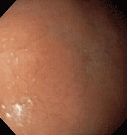
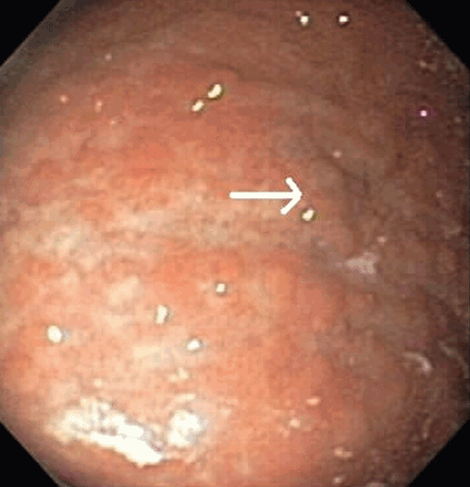
Due to recurrence of her marginal zone Bcell lymphoma of MALT type of the stomach, the patient is referred to an oncology service. She is treated with radiation, receiving 15 sessions of 30 Gy localized to the stomach. Three months after radiation therapy, she undergoes endoscopy again, which shows no evidence of the previously described nodules. Repeat biopsies are negative for H pylori and MALT lymphoma.

,

Annual surveillance endoscopy and computed tomography for the past 3 years have been negative for any tumor recurrence.