Role of MRI in breast cancer management


ABSTRACTIn breast cancer, different situations call for different imaging tests. Mammography is the test of choice for screening women with no signs or symptoms of breast cancer. For diagnosis, tailored mammographic views and ultrasonography are the norm. Magnetic resonance imaging (MRI) is highly sensitive for cancer staging, problem-solving, posttreatment surveillance, and other indications. It can detect primary breast cancers and additional foci of cancer that are occult to standard imaging. Continued improvements in technology and studies to assess outcomes will help to better define MRI’s role in breast cancer.
KEY POINTS
- Whether rates of death and local recurrence are reduced when additional breast tumors found by MRI are treated remains to be seen.
- MRI contrast enhancement occurs in many cancers, but it may occur for benign reasons; thus, the finding of contrast enhancement does not establish the diagnosis of breast cancer.
- The National Comprehensive Cancer Network currently recommends screening with both mammography and MRI starting at age 20 to 25 for women at high risk of hereditary breast cancer and ovarian cancer.
- A breast MRI evaluation costs about 10 times more than screening mammography and may not be covered by health insurance, but coverage for this indication appears to be improving gradually.
Problem solving
At times, mammographic findings are unclear as to whether a suspected lesion is truly present, often because of the radiographic density of fibroglandular breast tissue. In some cases, a lesion’s morphology is indeterminate for malignancy. These equivocal findings can usually be resolved with the combined use of tailored mammographic views, as noted above (eg, magnification or compression views), and directed ultrasonography.
If the findings are still inconclusive after this additional workup, MRI may be useful. Newer, improved MRI scanners can show structures as small as 0.5 mm, which helps the radiologist discern lesion morphology. Moreover, contrast-enhanced and temporally resolved imaging provides estimates of spatially localized enhancement patterns and kinetics, which in turn may offer clues as to whether a lesion is benign or malignant.
Screening patients at high risk
The National Comprehensive Cancer Network27 currently recommends screening with both mammography and MRI starting at age 20 to 25 for women at high risk for hereditary breast cancer and ovarian cancer. Risk factors include the following:
- A known BRCA1 or BRCA2 mutation in the patient or a family member
- A personal history of breast cancer, with two or more close blood relatives with breast or epithelial ovarian cancer at any age
- A close male blood relative with breast cancer
- A personal history of epithelial ovarian cancer
- Being in an ethnic group with a higher frequency of deleterious mutations (eg, Ashkenazi Jews)
- Mutations in p53 (Li-Fraumeni syndrome) or PTEN (Cowden syndrome).
MRI IN THE PREOPERATIVE EVALUATION: THE DEBATE
Numerous reports have shown that MRI can detect additional foci of breast cancer in a substantial number of women with a new diagnosis of breast cancer. While some argue that detecting these additional lesions should improve outcomes after the first operation and, hopefully, lead to lower rates of recurrence, the long-term consequences of MRI-directed changes in treatment have not been fully studied. Below is a summary of the arguments both against and for the use of breast MRI in staging.
The argument against preoperative MRI
Mastectomy was the routine treatment for breast cancer into the 1980s. The arrival of breast conservation surgery combined with radiation therapy offered major advantages with similarly low recurrence rates. Based on the results of controlled clinical trials with mortality as the end point, breast conservation therapy and mastectomy confer equivalent risk to the patient. Any increase in the rate of mastectomy prompted by MRI findings would represent a setback in the standard of care. And since radiation therapy is presumed to eradicate or delay progression of residual disease in most women who undergo conservation therapy, preoperative MRI would have little or no impact on rates of recurrence or death. Thus, MRI should not be used routinely in the workup of new breast cancers.28
The argument for preoperative MRI
The upper threshold amount of residual disease that can be eradicated by radiation therapy is not yet well established. There are as yet no MRI criteria for assessing the likelihood of standard treatment failure in individual patients with multifocal or multicentric disease, or with occult cancer in the contralateral breast. Although the rate of recurrence after breast conservation is low, it is not zero, and each patient should be offered the best possible chance for successful treatment. Detecting widespread disease can obviate inappropriate attempts at conservation, in which both lumpectomy with positive margins and re-excision with positive margins are carried out before the full extent of the disease burden is understood. Knowledge of the extent of disease at presentation will help the patient to make a more informed decision when presented with treatment options. A staging MRI examination showing only a single cancer lesion may permit the patient to choose conservation therapy with a high degree of confidence that no macroscopic disease will be missed at surgery.29
Challenges for future clinical trials
These issues will not be easy to resolve. Definitive answers can only come from controlled clinical trials with mortality as the end point, but for the data from these trials to be useful, the trials must use standardized MRI technique and interpretation criteria. Such standardization has yet to be accomplished.
In the absence of such guidance, it seems reasonable to use MRI for staging within the known limitations of the technique and with secure histologic confirmation whenever widespread disease is suspected from the MRI findings. In this way, the patient and her surgeon can select a treatment plan based on the most realistic assessment of disease burden.
CASE RESOLVED
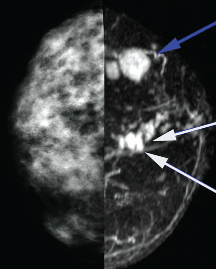
MRI to assess extent of disease (Figure 1) showed two enhancing lesions with irregular borders in the region of the proven cancer. The MRI enhancement kinetics of the lesions were consistent with malignancy. MRI also showed several additional, unsuspected, small, irregular lesions in the 12-o’clock region.
On the basis of these findings, a second ultrasonographic examination of the right breast was carried out, targeting the 12-o’clock region. One of the MRI-detected lesions was located, and biopsy showed invasive breast cancer of the same cell type as the palpable mass. With this evidence of multiple malignant lesions in the same breast, it was concluded that breast-conserving surgery would not be feasible. The patient underwent mastectomy with pathologic confirmation of the MRI findings.
Comment. This case demonstrates how breast MRI, when used appropriately, can lead to objective pathologic results that support the clinical decision to perform a mastectomy rather than breast conservation therapy.