Optic neuritis and risk of MS: Differential diagnosis and management


ABSTRACTOptic neuritis, a cause of sudden vision loss, often heralds the onset of multiple sclerosis (MS) within the next few years. It is important to distinguish optic neuritis from other types of optic neuropathy so that treatment can be started promptly, possibly delaying the onset of MS.
KEY POINTS
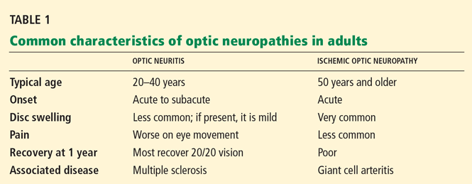
- Optic neuritis is most common in women in their 20s and 30s, whereas ischemic optic neuropathy, which is more common, primarily affects older people.
- The diagnosis of optic neuritis is primarily clinical, but magnetic resonance imaging confirms the diagnosis and, more importantly, assesses the risk of MS.
- Intravenous methylprednisolone (Solu-Medrol) does not affect the long-term visual outcome, but it speeds visual recovery and reduces the risk of MS. Surprisingly, oral prednisone seems to increase the risk of recurrent optic neuritis and is therefore contraindicated.
- Early treatment with interferon beta reduces the risk of MS and should be considered in patients at high risk.
CASE CONTINUED
Our patient undergoes a neurologic examination, which reveals an afferent pupillary defect in the right eye and visual acuity of 20/100 in the right eye and 20/20 in the left. Visual fields are normal in the left eye as assessed by confrontation, but there is a central scotoma in the right.
OTHER TYPES OF NEUROPATHY
The following should be included in the differential diagnosis of optic neuritis:
Ischemic optic neuropathy
Ischemic optic neuropathy is more common in patients age 50 and older, whereas optic neuritis is more common in younger patients. Most patients with ischemic optic neuropathy have hypertension, hypercholesterolemia, diabetes mellitus, obstructive sleep apnea, or other vascular risk factors. The disease has several important subtypes, as discussed below.

Although patients with nonarteritic anterior ischemic optic neuropathy typically have vasculopathic risk factors such as hypertension, diabetes mellitus, peripheral vascular disease, or hypercholesterolemia, there is no proven causation between the two. The age of these patients ranges from 50 to 70, with an average age of 66.
The disc appears swollen and may have flame or splinter hemorrhages (Figure 3). The cup of the involved disc is typically small. The visual loss is believed to be the result of poor perfusion in the circulation of the posterior ciliary artery, which supplies the optic nerve head.1 If the other eye also has a small cup, it is considered to be at risk of future ischemic events. In one study,14 the opposite eye became involved within the next 5 years in 14.7% of all cases. The risk of recurrent disease in the same eye is low (6.4% in another study15).
Arteritic anterior ischemic optic neuropathy is more common in patients over age 70 and is usually due to giant cell arteritis, which has a significant association with polymyalgia rheumatica. Patients may have jaw claudication, proximal myalgia and arthralgia, scalp tenderness, headache, fatigue, and a significantly elevated erythrocyte sedimentation rate and C-reactive protein level. These features should be looked for in the review of systems, although patients may not have all of them.
The funduscopic examination may reveal a pale, swollen disc, peripapillary hemorrhages, branch or central retinal artery occlusions, or cotton-wool spots.
Temporal artery biopsy is the gold standard for diagnosis, but treatment with corticosteroids should not be delayed pending biopsy or other test results.1
Thrombocytosis has been associated with a higher risk of permanent vision loss in patients with giant cell arteritis.16
Posterior ischemic optic neuropathy is the least common form of ischemic optic neuropathy. This diagnosis should be entertained in older patients who report acute, painless vision loss but have a normal funduscopic examination. Giant cell arteritis must be considered first in this setting.
Bilateral posterior ischemic optic neuropathy can occur (although rarely) in patients undergoing cardiac or spinal surgery.17 Risk factors thought to be associated with perioperative disease include anemia, hypotension, substantial blood loss during the surgery, surgeries longer than 6.5 hours, carotid atherosclerosis, and diabetes.18
There are no effective treatments for most ischemic optic neuropathies with the crucial exception of giant cell arteritis.
Neuromyelitis optica (Devic disease)
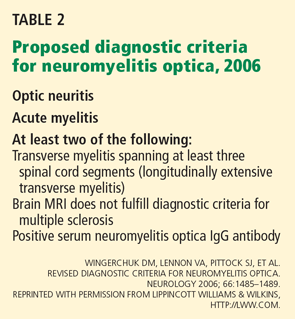
Neuromyelitis optica (Devic disease) is a combination of optic neuritis and transverse myelitis (Table 2). Clinically, the disease spares the nervous system except for the optic nerves and spinal cord. The onset of the optic neuritis may precede or follow the onset of the transverse myelitis by up to 2 to 4 years.19 Usually, the optic neuritis is bilateral and retrobulbar and results in severe vision loss, worse than that seen in patients with MS.19,20
The transverse myelitis may cause paraplegia or quadriplegia, depending on the location of the lesion in the spinal cord (cervical vs thoracic). The transverse myelitis in neuromyelitis optica is distinct from that seen in MS. In neuromyelitis optica, the transverse myelitis is longitudinally extensive, spanning more than three vertebral bodies in length. In MS, spinal cord lesions usually are more discrete and involve one or two spinal cord segments.21
Recently, serum neuromyelitis optica immunoglobulin G (IgG) antibody has been shown to be a significant biomarker of this disease. Its sensitivity ranges from approximately 60% to 70% and its specificity is greater than 90%.22 This antibody binds to aquaporin-4, an important water-channel protein in the blood-brain barrier of the central nervous system, and evidence suggests that it is involved in the pathogenesis of the disease.23
Initially, it was proposed that MRI of the brain had to be normal for neuromyelitis optica to be diagnosed.21 However, the proposed 2006 criteria allow for some abnormal T2 and fluid-attenuated inversion recovery (FLAIR) hyperintensities in the periaqueductal gray matter and diencephalon.22
The spinal fluid in neuromyelitis optica may show a pleocytosis larger than that seen in MS (> 50 white blood cells per mm3) and may have a significant neutrophilic component.21 Oligoclonal bands are not typically present.
It is still debated whether neuromyelitis optica is a separate disease from MS or a subset of it. The implications of this debate may affect its management, as discussed below.