Perioperative management of bariatric surgery patients: Focus on metabolic bone disease


ABSTRACTChronic vitamin D deficiency, inadequate calcium intake, and secondary hyperparathyroidism are common in obese individuals, placing them at risk for low bone mass and metabolic bone disease. After bariatric surgery, they are at even higher risk, owing to malabsorption and decreased oral intake. Meticulous preoperative screening, judicious use of vitamin and mineral supplements, addressing modifiable risk factors, and monitoring the absorption of key nutrients postoperatively are essential in preventing metabolic bone disease in bariatric surgery patients.
KEY POINTS
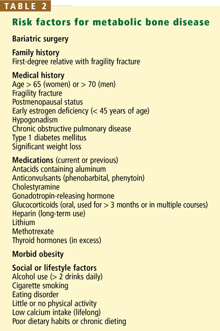
- Metabolic bone disease in obese patients is multifactorial: causes include sequestration of vitamin D in the adipocytes, inadequate nutrition due to chronic dieting, and lack of physical activity.
- Before bariatric surgery, one must look for and treat preexisting nutritional deficiencies.
- In the immediate postoperative period, aggressive strategies (ie, giving multivitamins and minerals intravenously and orally) can prevent nutritional deficiencies and secondary bone disease.
- Postoperatively, many bariatric patients require chewable or liquid supplements to facilitate adequate absorption.
- Clinical suspicion, timely interventions, and lifelong monitoring can prevent metabolic bone disease in bariatric surgery patients.
Calcium
Dietary calcium deficiency is a well-established risk factor for osteoporosis and fragility fractures. Therefore, supplemental calcium should be prescribed for patients who do not meet their defined need.34,35 Of note: a normal serum calcium level does not imply adequate calcium intake or absorption. Calcium homeostasis is tightly regulated and is maintained by a combination of gut absorption, bone resorption, and renal reabsorption. If dietary intake is inadequate, calcium is resorbed from the bone.
The duodenum is the major site of active calcium uptake, while the rest of the small intestine and the colon appear to absorb some calcium passively. When the physiologic need for calcium is increased, active transport appears to take place throughout the duodenum, the ileum, and, to a lesser degree, the jejunum and the colon.36
In the normal gastrointestinal tract, 20% to 60% of dietary calcium is absorbed.36–38 Patients who have lost absorptive surface area (eg, after Roux-en-y bariatric surgery) need to have their calcium intake optimized. However, optimal dosing based on the type of surgical procedure is currently undefined.
Judicious monitoring for compliance and adequate absorption is recommended. Some patients will stop taking their calcium supplement due to gastrointestinal side effects such as gas, bloating, or constipation. And for some patients, a standard calcium supplement may be insufficient to promote adequate calcium absorption. Measuring urinary calcium in a 24-hour sample can help in assessing the adequacy of calcium intake: abnormally low urine calcium in the presence of normal renal function suggests inadequate absorption. For patients reporting gastrointestinal side effects or those with a history of calcium oxalate renal stones, calcium citrate supplements are better tolerated, alter urine acidity, and often prevent further stone formation.
Vitamin B12 (cobalamin)
Vitamin B12 deficiency is associated with increased fracture risk, and it may be an important modifiable risk factor for osteoporosis.39–41 After surgery, malabsorption of vitamin B12 is commonly the result of altered gut function in the gastric pouch or sleeve, but malabsorption also occurs when more than 60 to 100 cm of terminal ileum has been bypassed.42
Vitamin B12 supplementation is recommended for all patients after bariatric surgery, because deficiency is common.42 Patients with relatively mild malabsorption can maintain their B12 level by taking 350 μg orally; however, many patients require lifelong subcutaneous injections.7,39,42–45
Magnesium
Magnesium appears to affect bone remodeling and strength, to have a positive association with hip bone mineral density, and to play an important role in calcium and bone metabolism.
Magnesium is absorbed in the distal small intestine by carrier-mediated and paracellular routes.46 When the distal small intestine is bypassed, magnesium deficiency occurs as a result of reduced absorption and chelation with unabsorbed fatty acids in the bowel lumen.42 Chronic hypomagnesemia impairs PTH secretion, resulting in altered calcium metabolism, hypocalcemia, and vitamin D abnormalities, further decreasing jejunal magnesium absorption.26,42,47
Few well-designed studies have investigated the effect of magnesium intake on bone health, and although there is evidence that postmenopausal women may benefit from magnesium supplementation, studies of magnesium supplementation after bariatric surgery are lacking.47,48
A prevailing misconception promoted by manufacturers of calcium-magnesium supplements and others is that magnesium is necessary for calcium absorption and efficacy. In fact, magnesium deficiency typically must be severe to impair calcium absorption. With usual dietary intake of magnesium and normal serum magnesium levels, no such relationship exists.49–53
THE ROLE OF DXA IN THE CARE OF THE BARIATRIC SURGERY PATIENT
DXA is the gold standard for measuring bone density. The results are reported as a T score and as a Z score.
The T score is the bone density in an area of interest expressed in standard deviations from the mean value of a reference database of young adults. The World Health Organization defines normal as a T score greater than or equal to –1, low bone mass (previously called osteopenia) as a score between –1 and –2.5, and osteoporosis as a score of less than or equal to –2.5. (If a fragility fracture has occurred, “established” or “severe” osteoporosis is present.) Of note: these criteria only apply to DXA of the posterior-anterior spine, femoral neck, and the proximal (33%) radius in post-menopausal women and men over the age of 50 years.54,55 The International Society of Clinical Densitometry has extended the criteria to include total hip measurements.56
The Z score should be used instead of the T score for premenopausal women and men younger than 50 years.56 The Z score is the patient’s bone mineral density expressed in standard deviations from the mean in a reference population matched for sex and age. A Z score greater than –2.0 is “within the expected range for age,” and –2.0 or lower is “below the expected range for age.” There are separate guidelines for DXA reporting in the diagnosis of metabolic bone disease in people younger than 20 years, and this topic is beyond the scope of this article.