A review of spinal arachnoid cysts


ABSTRACTThe symptoms of spinal arachnoid cysts are variable and nonspecific, so they are commonly misdiagnosed. Often the cysts are discovered incidentally on magnetic resonance imaging (MRI). If they cause no symptoms, no treatment is warranted regardless of the size of the cyst. Cysts that cause symptoms from mechanical compression of the spinal cord are best evaluated with MRI and surgically excised if possible.
KEY POINTS
- Spinal arachnoid cysts can occur at any age and at any spinal level.
- Symptoms vary widely but typically include waxing and waning pain and spastic or flaccid paraparesis.
- Most spinal arachnoid cysts are asymptomatic when diagnosed and are discovered incidentally on MRI or myelography.
- MRI and computed tomography help characterize spinal arachnoid cysts and differentiate them from abscesses and tumors.
- Symptomatic cysts should be surgically resected. If complete resection is impossible, fenestration of the cyst wall, drainage, or shunting may relieve symptoms.
- An asymptomatic spinal arachnoid cyst should be followed annually with serial imaging.
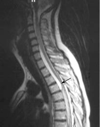
Imaging studies help evaluate pain from suspected nerve compression
Although most arachnoid cysts are found by MRI, it is inappropriate to initially order MRI to evaluate a cyst’s common presenting symptoms (eg, back pain, radiculopathy).
Plain radiography should be done first. Although arachnoid cysts are composed of fluid and soft tissue, which are not easily detectable on plain films, subtle and indirect signs of a chronic, large cyst may be visible.5
MRI is the next step if plain radiographs do not reveal bony abnormalities that could explain a patient’s symptoms.
Further studies help characterize the lesion
Diffusion-weighted MRI can help differentiate an epidermoid cyst from an arachnoid cyst. It may also help differentiate a cyst from an abscess or tumor: abscesses have areas of restricted diffusion, and tumors tend to lack cerebrospinal fluid signal in their central core. Diffusion-weighted MRI can also help evaluate spinal cord atrophy and inflammatory changes.1,6,12 If an arachnoid cyst accompanies a nerve root as it enters the neural foramen, this would also appear on MRI.
Myelography or computed tomographic (CT) myelography were used to further characterize the form and structure of spinal arachnoid cysts discovered on MRI in most reported cases, and most authors advocate these studies.1,3,8,12 Specifically, CT myelography has been used to look for a communication between the intraspinal subarachnoid space and the spinal arachnoid cyst, and it is sensitive in determining whether a communication exists, although it does not pinpoint the location of the communication very well.12 CT myelography is also invaluable for imaging the spine of patients who have contraindications to MRI.
Kinematic MRI (cine-MRI) is now widely available and can help evaluate for the presence of communications between the cyst and the subarachnoid space. Dural defects may be located by carefully scrutinizing cine-MRI images for pulsating turbulent flow voids, facilitating a more focused and minimally invasive treatment strategy.13
Neo et al12 used cine-MRI to evaluate and plan the surgical resection of a giant spinal extradural arachnoid cyst. MRI helped determine the initial diagnosis, and a pulsating turbulent flow void was observed by cine-MRI in the area later confirmed surgically to contain the communication between the cyst and the spinal subarachnoid space.
Cine-MRI is not necessary as part of the initial diagnostic evaluation for spinal arachnoid cysts. It is of particular value only to the surgeon, who can request it if needed.
HISTOPATHOLOGY
With hematoxylin and eosin staining, the walls of spinal arachnoid cysts are typically seen as fibrous and lined by meningothelial cells.
TREATMENT
Observe asymptomatic cysts
For incidentally discovered spinal arachnoid cysts that cause no symptoms—ie, most of them—surgery is not recommended. No correlation exists between the size of a cyst and the need for treatment. Yearly imaging should be done to detect any new abnormality and determine whether the cyst is truly benign.
If symptoms arise, reevaluation of the cyst with MRI should be immediately undertaken.
Remove symptomatic cyst if possible
For a patient with symptoms, treatment offers an excellent chance of neurologic recovery.
Aspiration of the cyst is not routinely advised. Although aspiration may intuitively seem like the best initial approach to management, it only temporarily improves symptoms. However, percutaneous aspiration under fluoroscopic guidance may be appropriate for determining whether a cyst is causing a patient’s symptoms and thereby predicting whether surgery can help. Surgery should be undertaken only after careful consideration, as postoperative complications, though uncommon, may be very troublesome for both the patient and the surgeon.
Complete resection is ideal treatment. The standard treatment of an isolated spinal arachnoid cyst is complete surgical removal of the cyst.1 Surgery typically results in excellent outcomes in terms of resolution of symptoms, and is effective across a large range of cyst sizes.
Drain cysts that cannot be resected. Unfortunately, not all isolated spinal arachnoid cysts can be fully resected, owing to their location or to intraoperative findings such as extensive adhesion of a cyst to the spinal cord. In such cases, fenestration of the cyst wall, percutaneous drainage, or shunting the cyst into the peritoneal cavity may relieve symptoms.1–3,6
Minimally invasive surgical techniques have also met with some success. Neo et al12 reported that they successfully treated a giant spinal extradural arachnoid cyst by selectively closing the dural defect with clips. Cine-MRI was used to pinpoint the communication, allowing for a focused, limited surgical approach requiring only fenestration. The dural surface of the cyst was examined with an operating microscope.
Endoscopic approaches have also been used to treat sacral extradural arachnoid cysts.7
SOME CASES ARE MORE COMPLEX
Managing spinal arachnoid cysts becomes more complex as cysts become more intricate in morphology and if multiple cysts exist across different vertebral levels. Surgical planning and intraoperative monitoring are also complicated if a spinal arachnoid cyst coexists with another central nervous system problem.
Cases have been reported of patients with coexisting spinal arachnoid cysts and lumbar disk herniation; in many, the latter problem was considered to be the cause of symptoms.7
Holly and Batzdorf3 described patients with both intradural arachnoid cysts and syringomyelia. Cysts were resected with the aid of an operating microscope, and intraoperative ultrasonography confirmed that normal pulsation of the subarachnoid cerebrospinal fluid had returned after resection. The syrinx cavities were not surgically manipulated, yet MRI taken 3 months after surgery revealed that they had significantly diminished in each case.
The best predictor of recovery in patients who undergo surgery for spinal arachnoid cysts is if the clinical presentation correlates with the defect.1,7 Usually the postsurgical prognosis is good, with significant to full neurologic recovery in patients with all cyst types and clinical presentations.