Dermatology for the pediatrician: Advances in diagnosis and treatment of common and not-so-common skin conditions
ABSTRACTAdvances have been made in understanding and treating both common and rare dermatologic conditions. Atopic dermatitis benefits from bathing and ceramide moisturizers. Common allergic contact dermatitis may have specific presentations. Tinea capitis is effectively treated with terbinafine. Infantile hemangiomas should be treated early in the disease course and respond well to propranolol; any white sign of ulceration should be noted. Localized alopecia areata responds well to topical clobetasol, avoiding the need for intralesional injections. Topical rapamycin can be used to treat tuberous sclerosis. Further understanding of genetics will help guide pediatricians to the proper diagnosis and treatment of skin conditions.
Car seats
Car seats also are associated with contact dermatitis but the specific allergen is unknown. The presentation is similar to toilet seat dermatitis as the posterior leg is affected while the anterior leg remains clear. This type of contact dermatitis is more frequently reported with the use of car seats made of tightly woven, shiny material. A cotton cloth barrier can be placed over the car seat to prevent skin contact.13
Sports equipment
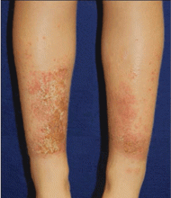
Allergic contact dermatitis may be associated with the use of shin guards and neoprene wetsuits, with p-tert butylphenol formaldehyde resin as the likely allergen.14 There is obvious delineation of the affected and nonaffected skin, which can help rule out atopic dermatitis (Figure 2). However, there is the potential for atopic dermatitis to develop with use of these items because the skin is occluded for long periods and will sweat, causing a reaction. Again, providing a barrier between the surface of the shin guard or wetsuit and the skin will help.
Metals
Finally, nickel and other metals are known sources of allergic contact dermatitis.15
TINEA CAPITIS
The challenge for this dermatologic condition is finding an effective treatment without a prolonged course of medicine. If tinea capitis continues for too long, the inflammatory process can result in permanent alopecia. The condition is caused by fungus, most commonly Trycophyton tonsurans.16 Terbinafine (3–8 mg/kg/day for 2–4 weeks) is superior to griseofulvin for T tonsurans; however, griseofulvin is superior to terbinafine for treating Microsporum species.17 Health insurance coverage of terbinafine is a concern for some families. Finally, children with tinea capitis should use a sporicidal shampoo, selenium sulfide, or ketoconazole to decrease the spread of spores.
INFANTILE HEMANGIOMAS
Propranolol
The major breakthrough in pediatric dermatology of the past decade has been the use of propranolol to treat infantile hemangiomas. Propranolol hydrochloride (Hemangeol) received US Food and Drug Administration (FDA) approval in March 2014 for the treatment of proliferating infantile hemangioma. The proposed mechanism of action is suppression of vascular endothelial growth factor and basic fibroblast growth factor in in vitro hemangioma-derived stem cells.18
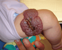
Propranolol is indicated for complex cutaneous, visceral, hepatic, and airway infantile hemangiomas. For most children, treatment with oral propranolol is preferable to prolonged treatment with systemic steroids. If propranolol is started early in the disease course, it can greatly reduce the need for extensive surgery, which is of particular importance when the nasal tip, oral mucosa, and face are affected. Children with large hemangiomas that are prone to ulceration also greatly benefit from propranolol treatment. Ulcerated hemangiomas are extremely painful and problematic, and can take months to heal (Figure 3).
Concomitant abnormalities and PHACE
A visceral evaluation is generally indicated for children with five or six lesions, but hemangioma size should also be considered when determining the need for additional tests. When a facial, scalp, or neck infantile hemangioma is larger than 5 cm, imaging studies are indicated to check for vascular anomalies in the brain and congenital heart defects (most commonly aortic problems).
A subgroup of these children have PHACE (posterior fossa anomalies, hemangioma, arterial lesions, cardiac abnormalities/aortic coarctation, and eye abnormalities) syndrome. A consensus guideline found that evidence supports treating these patients with propranolol.19 However, there have been isolated reports of acute ischemic stroke in PHACE syndrome patients on concomitant steroid therapy with severe arteriopathy. Therefore, before initiating propranolol therapy, infants with large facial hemangiomas who are at risk for PHACE should be evaluated with magnetic resonance angiography of the head and neck, and with cardiac imaging that includes the aortic arch.
Propranolol dosing
The FDA-approved dosing of propranolol hydrochloride is 1 to 3 mg/kg/day. Side effects include hypoglycemia, hypotension, exacerbation of asthma or respiratory infections, dental caries, cold extremities, and night terrors. These side effects should be closely monitored for, particularly in younger infants; however, propranolol has a relatively benign safety profile that should be taken into consideration in the risk/benefit analysis.
New pathogenesis insights
In addition to breakthrough treatment, a greater understanding of the pathogenesis of infantile hemangiomas has been attained. The most rapid lesion growth phase occurs at a younger age than was previously believed, and it is now thought to be greatest when patients are between 5.5 and 7.5 weeks old.20 Treatment initiation before the rapid growth period, rather than after, is crucial to successful outcomes.
Additionally, research has identified some of the clinical signs prior to ulceration of infantile hemangioma.21 A white central area occurring in a proliferating lesion is predictive of ulceration. This white coloration of the proliferation stage should not be mistaken for the graying of involution that occurs when infants are older than 3 months. Again, initiating early propranolol therapy should prevent some of the pain associated with hemangiomas that are likely to ulcerate.


