
CCND1-Negative Mantle Cell Lymphoma With Balanced CCND3-IGH Rearrangement: Case Report and Literature Review


Background: Mantle cell lymphoma (MCL) is characterized by the t(11;14)(q13;q32) translocation resulting in overexpression of cyclin D1 (CCND1). Rarely, MCL results from translocations involving other genes (ie, IGK, IGL, CCND2, CCND3, or CCNE). CCND1 immunohistochemistry and t(11;14) fluorescence in situ hybridization (FISH) may fail to detect cases with these alternative translocations.
Case Presentation: A former US Navy electrician who was aged 70 years with a history of metastatic urothelial carcinoma, papillary thyroid cancer, and smoking presented with asymptomatic thrombocytopenia, detected from routine laboratory assessment. The patient exhibited an abnormal karyotype, with a balanced translocation involving chromosomes 6 and 14, later confirmed by FISH to involve CCND3 in 6p21.1 and the immunoglobulin heavy chain (IGH) in 14q32. He was treated first with ibrutinib then zanubrutinib, but discontinued both due to significant bleeding. The patient’s performance status deteriorated after treatment with bendamustine + rituximab. He received palliative therapy for urothelial carcinoma before his death in hospice care.
Conclusions: We report a rare case of a CCND3-IGH–rearranged MCL, which presented a diagnostic challenge. This report highlights the use of complementary methods to diagnose the disease. A literature review of CCND1-negative MCLs reinforces the importance of an expedited diagnosis to optimize treatment.
Mantle cell lymphoma (MCL) accounts for 2.5% to 6.0% of non-Hodgkin lymphoma cases, with about 70% of all cases affecting men.1 MCL often occurs in middle-aged to older adults with a median age of onset of about 60 years.2 Of patients with MCL, 81.5% present with it in advanced stages (stage III or IV) with lymphadenopathy, hepatosplenomegaly, and bone marrow involvement. While variant morphologies may be present, the most common microscopic appearance is a monomorphic small to medium-sized lymphocytic proliferation.3 Immunophenotypically, these cells are uniformly positive for BCL2, mostly positive for CD5, CD43, and FMC7, and negative for CD10 and BCL6.4,5 CD23 is expressed variably among cases ranging from negative to weak positive. Expression of cyclin D1 (CCND1) is observed in > 95% of cases including the minority that are CD5 negative.6
This article describes a case of an older adult with CCND1-negative, t(11;14)-negative MCL. Cases of CCND1-negative MCL are rare. As of this writing, only 88 cases have been reported in the literature; this is the 14th case and fourth report involving CCND3, the second case with a CCND3-IGH rearrangement, the first with a noncryptic CCND3 rearrangement, and the first with a balanced t(6;14).
Case Presentation
A 70-year-old male with a history of hypertension, hyperlipidemia, coronary artery disease, chronic kidney disease, gout, asthma, benign prostatic hyperplasia, and bladder cancer presented with asymptomatic thrombocytopenia detected on routine laboratory assessment. A former US Navy electrician, the patient had a 60-pack-year smoking history. The patient’s family history included bladder cancer in his father and multiple unspecified family members with unspecified cancers. The patient’s urothelial carcinoma was diagnosed 4 years earlier as noninvasive papillary urothelial carcinoma and was treated with transurethral resection multiple times.
Workup of the patient’s thrombocytopenia included imaging, which revealed splenomegaly and retroperitoneal lymphadenopathy. A bone marrow biopsy revealed a normocellular marrow involved by an unclassifiable B-cell lymphoproliferative disorder.
The lymphoid neoplasm morphologically presented as rare small lymphoid aggregates composed of monomorphic small to medium-sized mature-appearing lymphocytes. Flow cytometry showed these cells comprised 12% of the cellularity and were positive for CD19, CD20 (strong intensity), CD23 (weak intensity), CD52, and FMC-7, with coexpression of CD5 (strong intensity) and monotypic expression of Λ surface light chains (strong intensity). Immunohistochemical staining showed the lymphoid aggregates to be negative for CCND1. A fluorescence in situ hybridization (FISH) assay using a CCND1/immunoglobulin heavy chain (IGH) fusion probe was negative. Other than the clinically insignificant findings of loss of chromosome Y in some of the metaphases and inv(9)(p11q13) in all metaphases, the karyotype was negative. Definitive classification of the lymphoproliferative disorder could not be made with the bone marrow specimen. A community care surgical consultation was placed to have a splenectomy performed. Outside pathology rendered a diagnosis of chronic lymphocytic leukemia (CLL)/small lymphocytic lymphoma based on the CD5 positivity, partial CD23 positivity, CCND1 immunohistochemistry (IHC) negativity, and negative CCND1/IGH FISH. Still asymptomatic, the patient was on active surveillance for the lymphoproliferative disorder.
Five years later, the patient was diagnosed with papillary thyroid carcinoma and underwent a total thyroidectomy. Three years following total thyroidectomy, incidental lymphadenopathy was detected on a lung cancer screening computed tomography; a subsequent positron emission tomography showed minimally hypermetabolic (max SUV 5.2) lymphadenopathy throughout the mediastinum, abdomen, and retroperitoneum. The patient reported no new physical restrictions, fevers, chills, recurrent infections, or night sweats when the incidental lymphadenopathy was detected.
Initial laboratory findings were significant for leukocytosis of 11.85 k/uL with lymphocytosis of 5.48 k/uL, Lactate dehydrogenase (LDH) was within normal limits (216 U/L). Fine-needle aspiration (FNA) of a 4R lymph node showed sheets of lymphoid cells of small size (Figure 1A). Flow cytometry found these cells to be positive for CD19, CD20 (moderate intensity), and FMC-7, with coexpression of CD5 (strong intensity) and monotypic expression of Λ surface light chains (strong intensity). Immunohistochemical staining showed them to be negative for CCND1 but positive for SOX11 (Figure 1B). Based on the CD5 positivity, CD23 negativity, FMC7 positivity, and SOX11 positivity, the care team considered MCL the likely, but unconfirmed, diagnosis (Figure 2). Still asymptomatic, the patient remained on active surveillance.
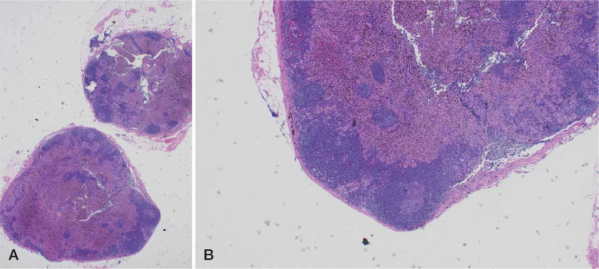
A, 2× magnification. B, 10× magnification.
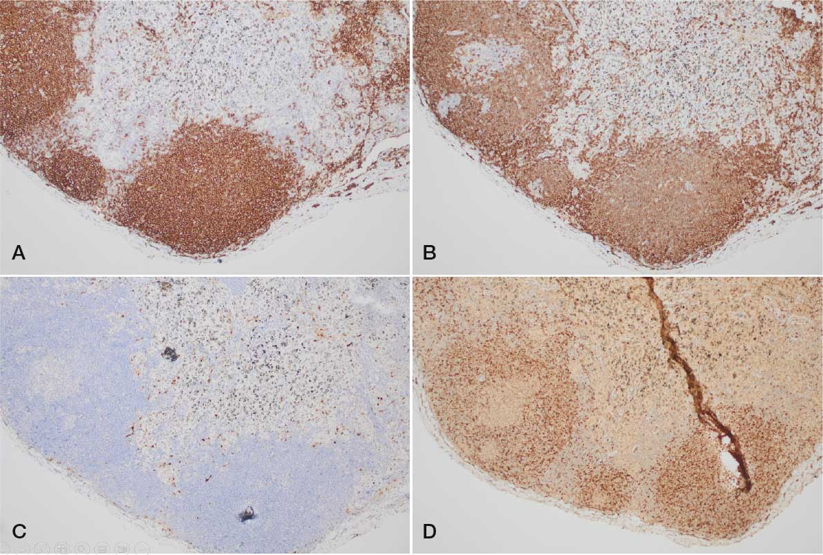
D, SOX11.
Two years later, the patient presented with dyspnea determined to be due to anemia; his complete blood count showed leukocytosis of 112.17 k/uL, absolute lymphocytosis of 44.54 k/uL, and thrombocytopenia of 91 k/uL. A bone marrow biopsy revealed hypercellular marrow (70% cellularity) with involvement by the known B-cell lymphoproliferative disorder as interstitial and nodular lymphoid infiltrates comprising 75% of the cellularity. The lymphoid cells were still monomorphic, appeared mature, and had no morphologic evidence of transformation. The immunohistochemical and flow cytometric findings were identical to those from the previous 4R lymph node. A FISH assay for t(11;14) was negative but FISH demonstrated an extra signal for IGH in 30% of cells, suggesting duplication of the 14q locus or an IGH rearrangement with another partner gene. The patient exhibited a complex and abnormal karyotype with 3 of 23 metaphase cells showing an apparently balanced translocation involving chromosomes 6 and 14, which are known to harbor the genes CCND3 in 6p21.1 and IGH in 14q32. A FISH study using a CCND3/IGH fusion probe confirmed this finding.
The complex karyotype showed the cells with t(6;14) to contain a mirror-image isochromosome 8q, leading to gain of 8q and loss of 8p, and an unbalanced rearrangement of 12p. A neoplasms targeted gene panel by next-generation sequencing was performed and revealed pathologic variants of ATM (p.K2756), TET2 (p.R1516), SRSF2 (p.P95L), and ASXL1 (p.Q708). A FoundationOne Liquid CDx assay showed the following pathogenic variants: ARID1A E1779*, ASXL1 Q708*, ATM K2756*, ATM V1569fs*33, FGFR3 R248C, NF1 A1785fs*9, PIK3CA E542K, SMARCA4 R1189Q, STAG2 S320fs*2, STAG2 splice site 2097-56_2142del102, and TET2 R1516*. The assay also showed the following variants of uncertain significance: ARID1A E1802Q, ATM G2063V, BAP1 R701L, CD79A T140N, CHEK2 A537T, GRM3 amplification, HGF amplification, KDM6A Q1212P, KIT V399I, KLHL6 L90H, MEF2B R53C, MST1R A1065T, NTRK1 G18E, PDGFRB E162K, PIK3C2B S1041T, PPARG R212Q, SMARCA4 S813L, TEK N1018S, TERT promoter -124C>A. The ATM K2756* variant was flagged as a potential germline variant with a 30.3% variant allele fraction (VAF). Overall tumor mutational burden was 15 mutations per megabase and there was no evidence of microsatellite instability.
The patient was treated with ibrutinib but it was discontinued after he experienced significant bleeding due to the anticoagulant effect of Bruton tyrosine kinase inhibitors.7 He was switched to zanubrutinib and again experienced bleeding that necessitated discontinuation. The patient then completed a cycle of bendamustine + rituximab, but chemotherapy was discontinued due to deteriorating performance status.
Following discontinuation of therapies, an L3 lesion was detected on imaging and determined to be a metastasis of the patient’s urothelial carcinoma that had formerly been confined to the lower urogenital tract with recurrences treated by transurethral resection and maintained by gemcitabine after bacille Calmette-Guérin. For the metastatic urothelial carcinoma, he completed palliative radiation. Due to the patient’s deteriorating state, he was not a candidate for chemotherapy/radiotherapy, entered hospice care, and died shortly thereafter.
Discussion
Dysregulation of oncogenes by translocation in juxtaposition to regulatory elements of the transcriptionally active IGH (14q32) or Ig light chain (Κ 2p11 [IGK] or Λ 22q11 [IGL]) genes is key in the pathogenesis of MCL. FISH studies have demonstrated that a translocation involving IGH (14q32) and CCND1 (11q13.3) is the major chromosomal abnormality of MCL seen in > 95% of cases.8,9 Occasionally, strong immunohistochemical positivity for CCND1 is not supported by FISH studies, and cryptic rearrangements of IGK or IGL enhancers with CCND1 in some of these cases give rise to CCND1 overexpression.10 Less frequently, an alternative cyclin activation is caused mostly by IGH and CCND2 fusions or even more rarely by fusions involving CCND3 or CCNE. Small scale research found that secondary molecular alterations in these groups were similar to CCND1-positive cases, highlighting gains of 3q, 8q, and 15q, and losses of 1p, 8p23- pter, 9p21-pter, 11q21–q23, and 13q.11
A literature review found 23 articles that described 84 cases of CCND1CCND1-negative MCL. Of the 84 cases, 54 (64%) exhibited CCND2 alterations (6 translocations with IGH, 25 translocations with IGK, 7 translocations with IGL, and 12 cases without an identifiable partner), 13 (15%) exhibited CCND3 alterations (2 translocations with IGH, 8 translocations with IGK, 2 with IGL, and 1 case without an identifiable partner), 5 (6%) exhibited CCNE alterations, and 12 (14%) cases were not further classified (Table).12-24 The reported cases included 62 males and 22 females with a mean age of 64.12 years and no statistically significant difference between sexes.
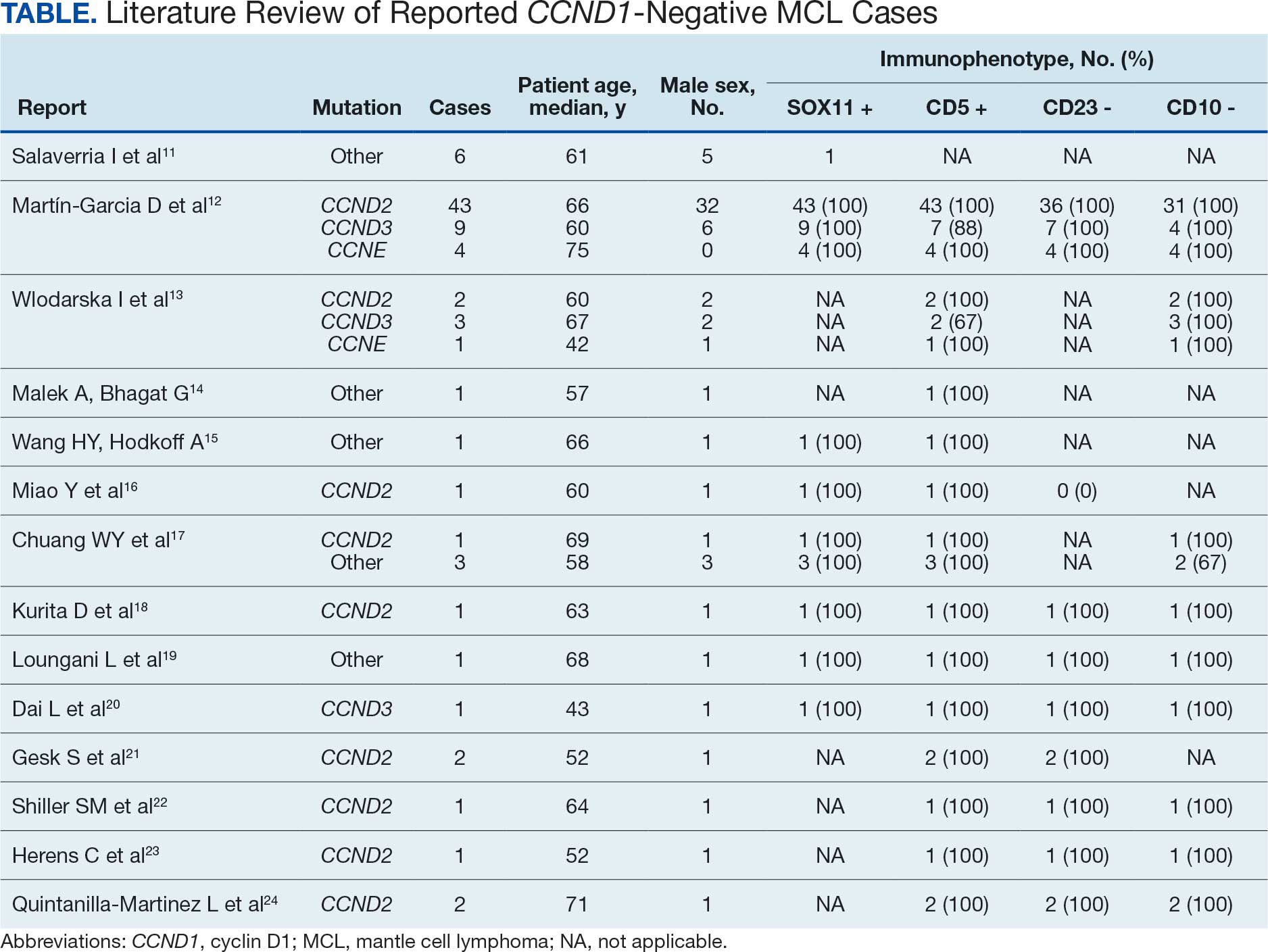
The overall survival and prognosis of CCND1-negative MCL are not well established due to the rarity of the condition, yet evidence suggests outcomes are similar to those of conventional MCL. Immunohistochemically, cases were CD20-positive, and reviewing the literature, the rate of SOX-11 positivity stood at 100% (66/66) wherever it was reported and was noted to be absent from older works. CD5 was positive in 97% of cases (76/78) and CD10 was negative in 98% of cases (56/57). Considering the high concordance between cases, no statistical difference was noted between sexes or underlying mutation.
The case outlined in this article showed similar findings with positivity for CD19, SOX- 11, FMC-7 and CD20, and coexpression of CD5. CCND3 rearrangement has been demonstrated in CLL; however, both the immunophenotype and morphological appearance of this case provide overwhelming evidence to support an MCL diagnosis.25
Conclusions
This case highlights nontraditional rearrangements in MCL and the use of corroborative complementary methods in the diagnosis of MCL. Considerable advances in the understanding of the molecular pathogenesis of CCND1-negative MCL have been made, yet the rarity necessitates additional reports of cases such as this one to further understand the range and frequencies of genetic abnormalities. These advances should expedite diagnosis and optimize management.
