Authors’ Disclosure Statement: The authors report no actual or potential conflict of interest in relation to this article.
Dr. Fitzpatrick is Assistant Professor of Radiology, Department of Radiology, Icahn School of Medicine at Mount Sinai, New York, New York; Director, Mount Sinai Queens Imaging, Astoria, New York; and Radiology Site Director, Mount Sinai Brooklyn Heights, Brooklyn, New York. Dr. Menashe is a Fellow, Department of Radiology, Montefiore Medical Center, Bronx, New York.
Address correspondence to: Darren Fitzpatrick, MD, Department of Radiology, Mount Sinai Queens; 25-10 30th Avenue, Astoria, NY 11102 (email, darren.fitzpatrick@mountsinai.org).
Am J Orthop. 2018;47(5). Copyright Frontline Medical Communications Inc. 2018. All rights reserved.
Darren Fitzpatrick, MD Leo Menashe, MD . Magnetic Resonance Imaging Evaluation of the Distal Biceps Tendon . Am J Orthop.
May 23, 2018
TAKE-HOME POINTS
There are a variety of injuries to the distal biceps tendon.
Injuries vary from tendinosis to full thickness, retracted tears.
The degree of retraction of full thickness tears depends on the integrity of the lacertus fibrosis.
The FABS view allows for MRI of the entire length of the distal biceps tendon.
MRI is the most useful imaging modality to determine the integrity of the postoperative biceps tendon.
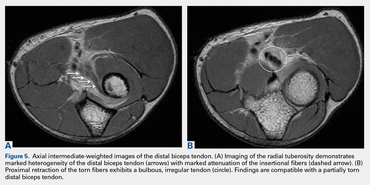
Partial distal bicep tears are characterized on MRI by focal or partial detachment of the tendon at the radial tuberosity with fluid filling the site of the tear. The degree of partial tearing can be assessed on MRI (Figures 5A, 5B).
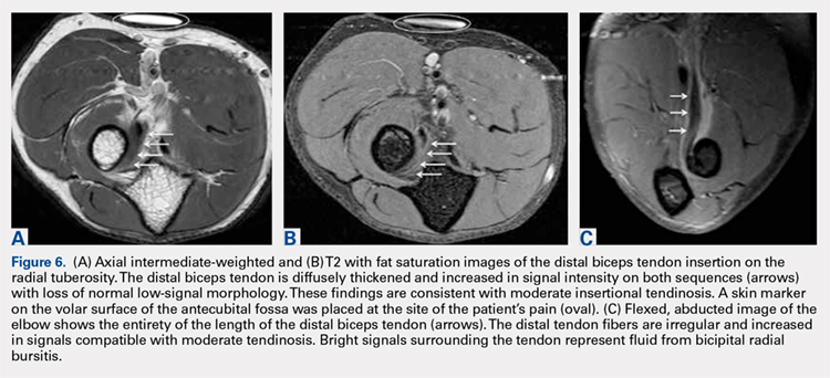
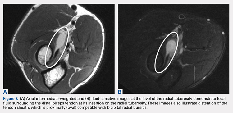
In distal biceps tendinosis, increased signals of thickened tendon fibers at the radial tuberosity are evident without focal discontinuity7,8 (Figures 6A-6C). Patients may display attenuation of the distal tendon fibers or adjacent fluid distension representing bicipitoradial bursitis (Figures 7A, 7B).
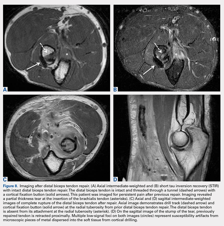
MRI is useful in assessing the distal biceps tendon in the postoperative setting to evaluate the integrity of a repaired tendon. Cortical fixation button technique for repair creates minimal susceptibility artifacts on MRI. Postoperative MRI typically demonstrates a transverse hole drilled through the proximal radius at the site of the tuberosity with a cortical fixation button flush against the posterior radial cortex (Figures 8A-8D).
The postoperative complication of heterotopic ossification can occasionally be observed on plain radiograph at the site of surgery, but it is less common with the current surgical technique than in the past.11