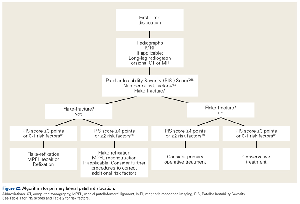
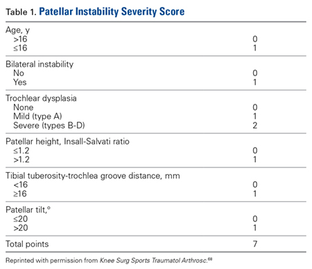
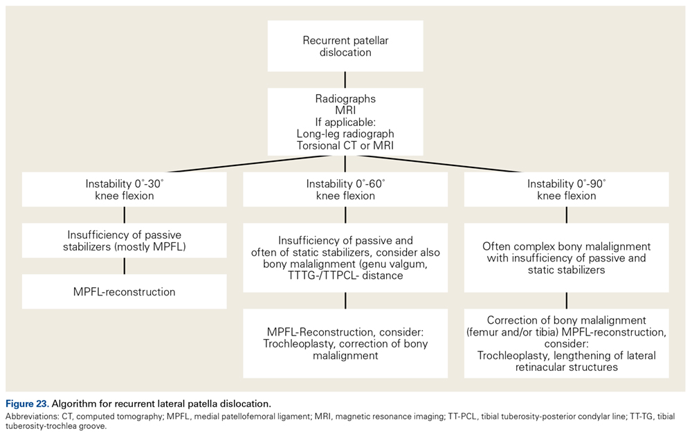
The incidence of lateral patella dislocations is high, particularly in young females. Beside traumatic cases, many patients present with specific anatomical factors that predispose to lateral patella dislocations (torsional abnormalities of the femur or the tibia, trochlea dysplasia, patella alta, etc). It is of utmost importance to correct those pathologic factors during concomitant procedures as isolated reconstructions of the medial patellofemoral ligament would fail in the presence of severe anatomic risk factors. This article provides a comprehensive instruction on how to analyze the risk factors for lateral patella dislocation (anatomy, physical examination, imaging) and reports the authors’ favorite surgical techniques. Moreover, treatment algorithms are provided for primary and recurrent cases of lateral patella dislocation.
Imaging
Radiographs are the basis for each patient’s imaging analysis. For a patient with valgus or varus clinical appearance, a weight-bearing whole-leg radiograph is used to precisely assess the degree of deformity in the frontal plane. A true lateral radiograph (congruent posterior condyles) provides information about patellar height (patella alta/infera). Most indices that quantify patellar height use the tibia as reference (eg, tuberosity, anterior aspect of articulation surface).
The Caton-Deschamps index measures the length of the articulating patella surface (A) and the distance from the most distal point of the patellar surface to the most anterior aspect of the articulating surface of the tibia (B); distance B divided by distance A yields the index, with values >1.2 indicating patella alta and values <0.6 indicating patella infera34 (Figure 8).
The lateral radiograph should also be checked for trochlear dysplasia, indicated by the crossing sign, the trochlear bump, or both (Figure 9). A weight-bearing anteroposterior (eg, Schuss) radiograph, which provides information on accompanying degeneration of the tibiofemoral joint, should be performed, particularly for elderly patients.
MRI is the gold standard for LPD diagnosis—it can be used to easily identify soft-tissue lesions and establish their patellar or femoral location (eg, MPFL rupture). MRI also provides information on potential pathologies of quadriceps tendon, patella tendon, and infrapatellar fat pad. Compared with radiographs, MRI is more sensitive in detecting osteochondral lesions in LPD.
Furthermore, functional measurements (eg, patellar tilt, patellar shift) can be made on axial MRIs, as the posterior condyles provide a proper reference line. MRI also plays a key role in determining accompanying degenerative changes in patients with LPD and therefore helps distinguish between joint-preserving and prosthetic procedures.
MRI also provides information on patellar height. In contrast to the radiographic patellar height assessment mentioned earlier, the patellotrochlear index of Biedert and Albrecht35 allows patellar height to be related to the proximal end of the trochlea.
From a biomechanical point of view, it seems more appropriate to determine patellar height respective of the trochlea, the articulating partner of the patella.
Further typical imaging parameters in LPD—such as TT-TG distance, femoral and tibial torsion values, and Dejour trochlear dysplasia—are also reliably shown with MRI. With lateral radiographs, MRI classifies trochlear dysplasia as type A (flatter than normal, with sulcus angle >145°), type B (flat), type C (convex), or type D (convex with supratrochlear spur and cliff) (Figures 10A-10D).
Treatment
MPFL Reconstruction
Isolated MPFL reconstruction is commonly regarded as a standard, straightforward procedure.
However, some authors have reported a considerable complication rate.36 Most failures have been attributed to technical errors and inappropriate indications.
The indication for isolated MPFL is regarded as inappropriate in patients with coexisting severe osseous pathologies, such as high-grade trochlear dysplasia and pathologic TT-TG distance.37,38
We recommend against performing isolated MPFL reconstruction in patients with any of these conditions: TT-TG distance >20 mm; femoral anteversion >30°; type C or D trochlear dysplasia; severe patella alta; advanced patellofemoral cartilage degeneration; or tibiofemoral valgus >5°.
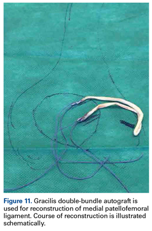
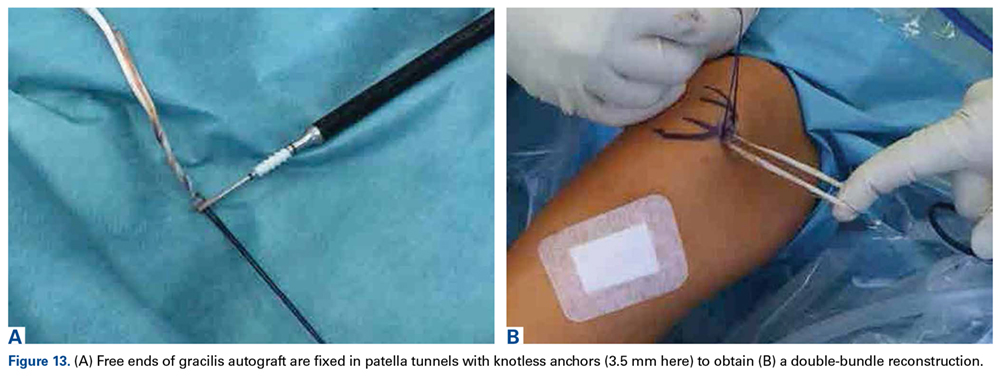
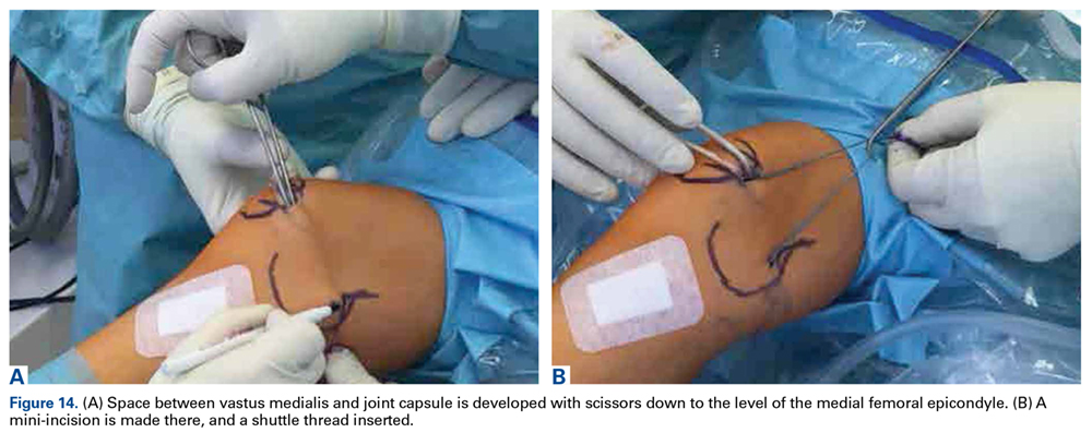
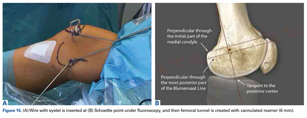
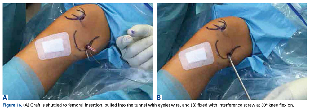
With use of accurate indications and surgical technique, isolated MPFL reconstruction provides good outcomes in patients with LPD.39,40 MPFL reconstruction has been performed with a wide variety of surgical techniques (eg, graft type, single-bundle vs double-bundle, fixation type). Our preferred technique (double-bundle gracilis autograft with aperture fixation) is detailed in Figures 11 to 16.
Trochleoplasty
In cases of recurrent LPD or a flat or convex trochlea (Dejour type B, C, or D dysplasia), deepening trochleoplasty should be considered.
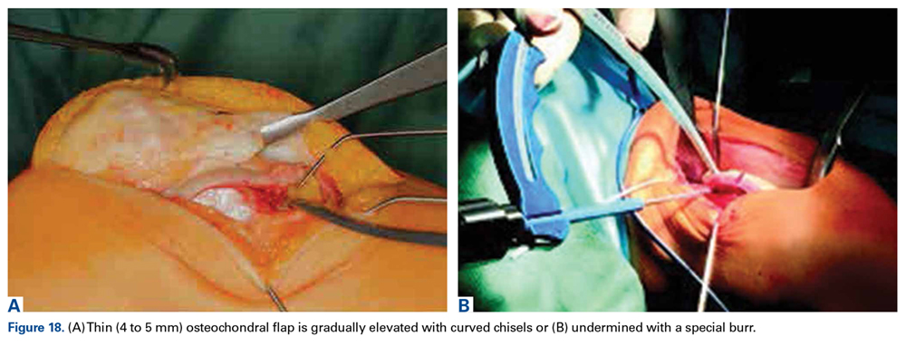
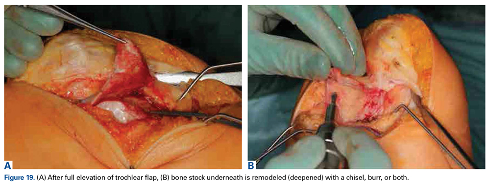
Trochleoplasty is performed to reduce too prominent anterior bone stock and to increase conformity with the patella (concave groove), and to create a lateral trochlea facet as restraint against lateralizing quadriceps pull. Many authors have reported good clinical outcomes of trochleoplasty in patients with LPD caused by a dysplastic femoral trochlea.41-48
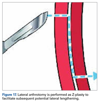
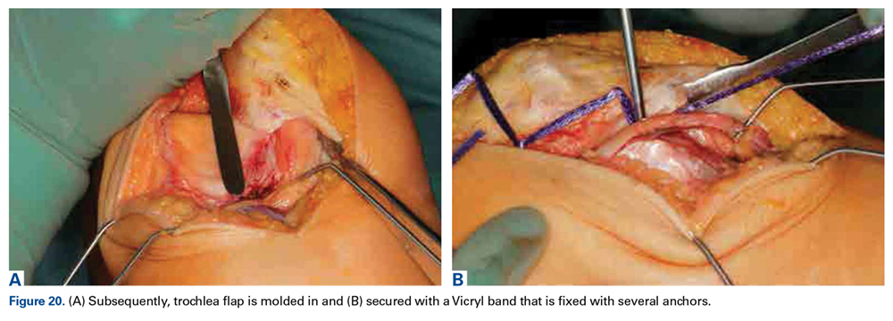
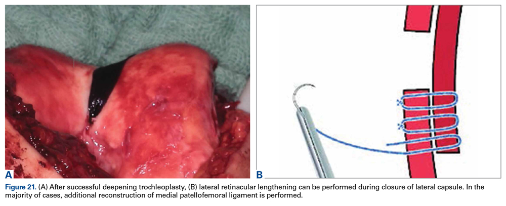
In many cases, MPFL reconstruction is added to trochleoplasty. Several authors have recommended against performing trochleoplasty in cases of open physis,49-52 which makes treatment of LPD in skeletally immature patients a special challenge, as trochlear dysplasia is often the key factor in failure of alternative procedures in the young.51 Another contraindication to trochleoplasty is severe cartilage degeneration. Our preferred surgical technique is described in detail in Figures 17 to 21.
Osteotomy
The most popular type of osteotomy in the setting of LPD is the transfer of the TT (TTT).
Many authors have reported good clinical outcomes with medializing TTT in patients with LPD and large TT-TG distances.53-57 Similarly, good outcomes have been found with distalizing TTT in patients with LPD and patella alta.58,59 We suggest routinely combining distal or medial TTT with MPFL reconstruction.60 TTT can be tailored to the patient’s pathology by combining medialization and distalization. Our preferred technique is to medialize the tuberosity so it ends with a TT-TG distance of at least 10 mm (avoid overcorrection).