Combined Anterior-Posterior Decompression and Fusion for Cervical Spondylotic Myelopathy


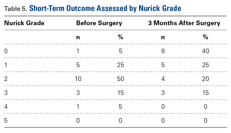
We conducted a study to evaluate the operative details, perioperative complications, and short-term outcomes associated with combined anterior-posterior decompression and fusion (CAPDF) for treating cervical spondylotic myelopathy (CSM). We retrospectively reviewed the charts of 21 patients who underwent CAPDF at our institution. Pertinent information, including demographics, surgery indication, perioperative complications, operative time, levels fused (and number of levels fused) anteriorly and posteriorly, estimated blood loss, and length of stay, was gathered. Outpatient follow-up data were available for 20 of the 21 patients, and postoperative neurologic status was evaluated with Nurick grades as well as by subjective means. Mean age was 62.1 years (range, 44-79 years). Of the 21 patients, 9 were female, and 12 were male. Before surgery, all patients had a diagnosis of CSM of varying degree. Mean number of levels fused was 2 (range, 1-3) anteriorly and 3 (range, 1-4) posteriorly. Mean operative time, which included patient repositioning, was 4 hours 55 minutes (range, 3:04-6:22). Mean estimated blood loss was 131 mL (range, 55-278 mL), and mean length of stay was 5 days (range, 2-10 days). The most commonly encountered complication was dysphagia (28.6%, 6/21). Neither neurologic instability nor mortality was observed after surgery. Neurologic status was subjectively improved for 19 patients and unimproved for 1 patient; no patient’s neurologic status was worse. Mean Nurick grade was 1.9 before surgery and 1.1 after surgery (mean difference, 0.80; P < .001), at a mean follow-up of 96 days (range, 51-149 days). When indicated, CAPDF is an efficient and effective treatment for CSM. This study found the procedure to be associated with minor complications, no new neurologic deficits, and high levels of neurologic improvement. The positive short-term outcomes and low rate of long-term complications in our study, combined with data from previous comparative studies, suggest that same-day surgery is superior to staged surgery.
Operative Technique: Posterior Approach
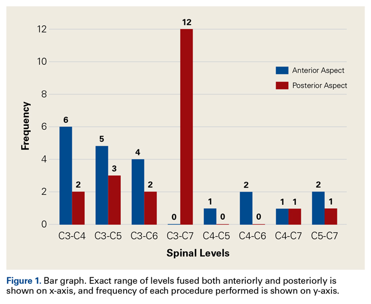
A midline incision was made through the skin and subcutaneous tissue to the level of the deep cervical fascia. Then, dissection was performed to the tips of the lateral masses. Instrumentation and fusion preceded spinal decompression. This order, chosen to preserve bony landmarks for guidance during instrumentation, did not interfere with subsequent decompression. Segmental spinal instrumentation was placed using lateral mass screw-rod fixation. After the laminae and ligamenta flava were bilaterally mobilized, the entire bony ligamentous complex spanning the area of fusion was removed en masse (most commonly C3–C7) in order to decrease the number of instrument passes near the spinal cord. Next, a modest foraminotomy was performed to extend the opening laterally and ensure adequate decompression of the nerve roots. Autograft harvested from the spinous processes and laminae was used. The posterior portion of the operation contributed significantly to blood loss and postoperative pain during the perioperative period. We recommend performing a very meticulous dissection to minimize these consequences. No patient in this study required a halo orthosis.
Results
Twenty-one patients with CSM were treated with CAPDF between February 2010 and March 2015 (Table 1). 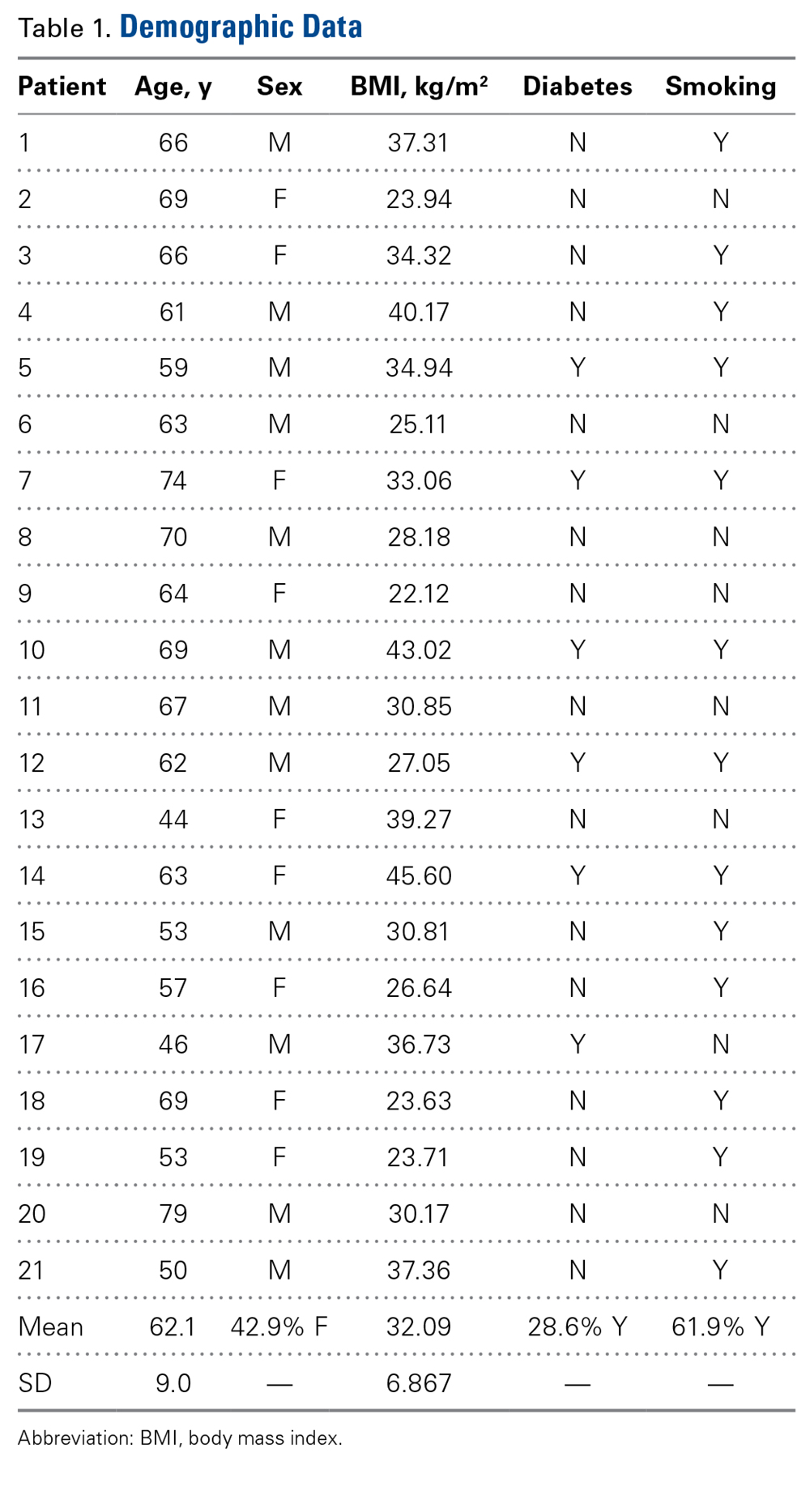
Table 2 summarizes the operative data. Mean number of levels fused was 2 (range, 1-3) anteriorly and 3 (range, 1-4) posteriorly. 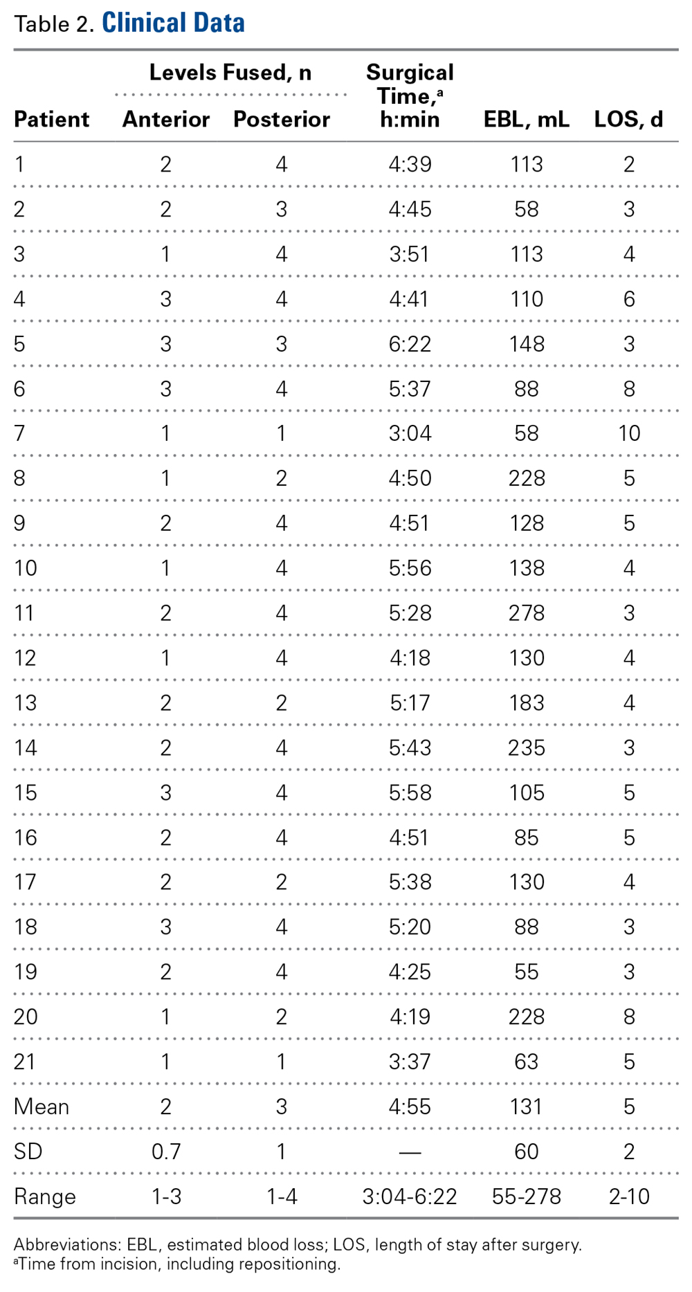
Of the 21 patients, 9 (42.3%) had at least 1 complication during the perioperative period. Table 3 summarizes all encountered complications. Neither neurologic instability nor mortality was observed after surgery. 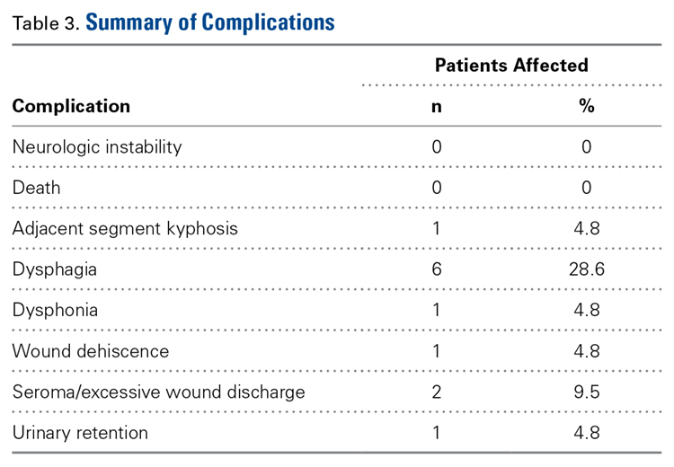
Patient 7 was lost to follow-up. For the other 20 patients, mean time to “3-month follow-up” was 96 days (range, 51-149 days). The most commonly noted improvements in quality of life included resolution of numbness, improvement in gait, and return to previous activities, such as walking and even exercising. 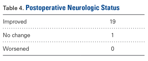
Representative Case
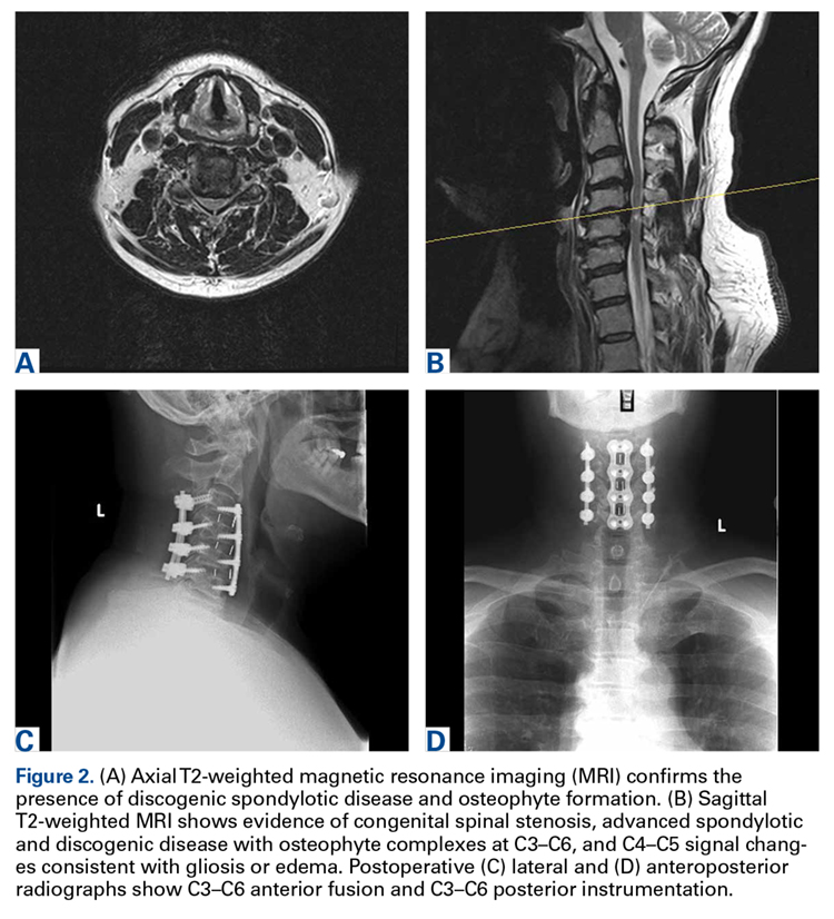
Patient 15, a 53-year-old man, presented with complaints of dysesthesias of the hands. Focused neurologic evaluation at the time revealed limited CS range of motion on extension. The patient (Figures 2A-2D) was diffusely hyperreflexic and had pathologic spread in the upper extremities.