Acute Shortening Versus Bridging Plate for Highly Comminuted Olecranon Fractures


Severely comminuted olecranon fractures, for which elbow stability becomes the main goal of surgery, remain a challenge for surgeons.
We conducted a study to determine the percentage loss of articular surface at which a bridge plating (BP) construct becomes too unstable and an acute shortening (AS) construct is required. The olecranon process of 8 fresh-frozen cadaveric upper limbs was serially resected. At each resection, the simulated fracture was fixed first with BP and then with AS. Stability was tested by performing valgus and varus stress tests at various angles under fluoroscopy.
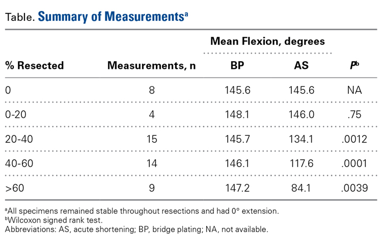
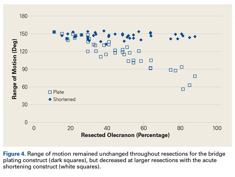
As many as 6 serial resections were made on the cadaveric models. Maximum resection was 88%. The ulnohumeral joint remained stable to valgus and varus stress at all resections for both BP and AS. The elbow joint lost a significant amount of flexion with AS above 20% resection.
The ulnohumeral joint can tolerate substantial loss of articular surface in the olecranon before becoming unstable. In this study, range of motion was preserved more with the BP construct than with the AS construct. The presented data may be considered when approaching a severely comminuted olecranon fracture in which the articular surface cannot be reconstructed.
Analysis
ImageJ software was used to analyze the C-arm radiographs. Measurements were divided into 4 groups of joint surface loss caused by the resections: 0% to 20%, 20% to 40%, 40% to 60%, and >60%. Differences in ROM between the BP and AS constructs were analyzed with a Wilcoxon signed rank test with statistical significance set at P < .05 (Prism 6; GraphPad Software).
Results
As many as 6 serial resections were made before the proximal fragment of the olecranon was judged too small to be secured to a plate with at least 2 screws. Only 7 specimens were large enough for the fifth cut, and only 4 were large enough for the sixth cut. After the final resection, mean loss of olecranon length was 77.3% (range, 63.7%-88%; median, 80.6%). All elbow specimens remained stable to manual valgus and varus testing in full extension, 30° of flexion, and full flexion in both supination and pronation. There was no medial or lateral opening of the ulnohumeral joint on fluoroscopy throughout testing, for either the BP or the AS constructs. There was no anterior or posterior subluxation throughout the entire ROM.